

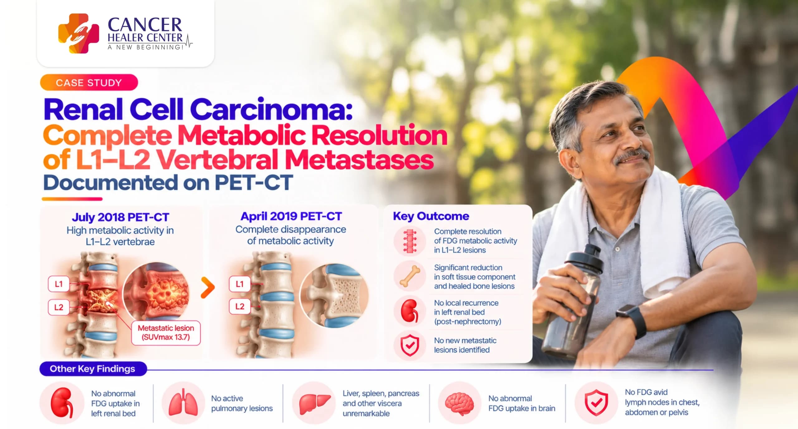
A Strong Response Documented Across Follow-Up Scans
This case study documents the clinical progress and therapeutic response of a 59-year-old male with Renal Cell Carcinoma (RCC) status post-left nephrectomy who developed spinal metastasis. Sequential evaluation using whole-body 18F-FDG PET-CT scans demonstrated a remarkable transition from high-grade metabolic activity in the L1–L2 lumbar vertebrae to complete metabolic disappearance and lesion healing following targeted therapy.
Mohan Bhosale’s Case at a Glance
| Patient Name: | Mr. Mohan Bhosale |
| Age / Gender: | 59–60 Years / Male |
| Referred By: | Dr. Sachin Almel / Dr. Sachin V. Almel |
| Primary Diagnosis: | Renal Cell Carcinoma (Left Nephrectomy, May 2016) |
| Metastatic Site: | Posterior aspect of L1 and L2 lumbar vertebrae extending into the spinal canal |
| Treatment Phase: | Oral targeted chemotherapy / Immunotherapy |
| Primary Outcome: | Complete resolution of FDG metabolic activity in L1–L2 spinal lesions and size reduction of soft tissue components |
Mohan Bhosale’s Treatment Journey
Mr. Mohan Bhosale underwent a left radical nephrectomy in May 2016 for renal cell carcinoma. In July 2018, he presented with lower back pain and lower limb weakness. Diagnostic imaging revealed a solitary metastatic lesion involving the L1–L2 vertebrae with spinal canal extension. Following the start of oral therapy in early 2019, follow-up imaging confirmed structural stabilization and complete disappearance of active metabolic disease.
What the July 2018 PET-CT Showed
A baseline 18F-FDG PET-CT scan performed at Jaslok Hospital & Research Centre on 30-Jul-2018 (Dose: 4.8 mCi 18F-FDG) evaluated the cause of spinal symptoms.
High-Grade FDG-Avid L1–L2 Vertebral Lesion
- Metabolic Activity: Intensely increased FDG uptake (SUVmax = 13.7) noted in a hypodense soft tissue lesion in the posterior aspect of the L1 and L2 lumbar vertebrae.
- Bone Involvement: Destruction of the spinous process and inferior facets of the L1 vertebra with erosion of the L1–L2 facet joint.
- Spinal Extension: The soft tissue component extended directly into the spinal canal.
No Local Recurrence in Left Renal Bed
- Operative Site: No abnormal increased FDG uptake or soft tissue mass was identified in the left nephrectomy bed.
Other Organs Assessed
- Viscera & Organs: Brain, lungs, mediastinum, liver, spleen, pancreas, gallbladder, right kidney, and adrenals were completely clear of abnormal tracer uptake or metastasis.
- Lymph Nodes: No significantly enlarged or FDG-avid lymph nodes were observed in the chest, abdomen, or pelvis.
How the July 2018 Scan Was Interpreted
- Impression: High-grade metabolic activity in the posterior aspect of L1 and L2 lumbar vertebrae causing bone destruction and canal extension, highly suspicious for metastatic disease.
- Local Site: No evidence of local tumor recurrence in the left renal bed or distant visceral metastasis elsewhere in the body.
What the April 2019 PET-CT Confirmed
A follow-up whole-body 18F-FDG PET-CT scan was performed at Jeeyo PET CT & Nuclear Imaging Centre on 23-Apr-2019 (Dose: 5.4 mCi 18F-FDG).
Complete Disappearance of Metabolic Activity at L1–L2
- FDG Avidity: The previously hypermetabolic L1–L2 spinal lesions became completely FDG non-avid.
- Metabolic Response: Complete clearance of abnormally elevated glucose uptake (SUVmax normalized).
Significant Reduction in Soft Tissue Lesion Size
- Morphological Response: Significant reduction in the size of the soft tissue component surrounding the L1–L2 vertebrae compared to the July 2018 scan.
Surgical Fixation and Healed Lytic Lesions
- Surgical Hardware: Spinal fixation screws were noted involving the D11–L3 vertebrae.
- Bone Status: Lytic lesions at L1–L2 showed imaging signs consistent with healed disease.
No Other Active Lesions Identified
- Whole-Body Survey: No tracer-avid lesions identified anywhere in the rest of the skeleton, brain, thorax, abdomen, or pelvic cavity.
- Renal Bed: Sustained clearance of disease in the left renal bed.
The Response Documented Over Time
| [May 2016] Left Radical Nephrectomy for Renal Cell Carcinoma │ ▼ [Jul 2018] PET-CT: High-Grade SUVmax 13.7 FDG-Avid L1–L2 Spinal Metastasis │ ▼ [Feb 2019] Initiation of Targeted Oral Systemic Therapy │ ▼ [Apr 2019] Follow-up PET-CT: Complete Disappearance of FDG Activity; Healed L1–L2 Lesions |
What the Two Reports Confirm
- Complete Local Metabolic Remission: Conversion of an intensely active metastatic spine lesion (SUVmax = 13.7) to non-avid, metabolically inactive tissue.
- Structural Disease Control: Reduction in soft tissue bulk and stabilization of lytic bone defects following spinal fixation.
- Absence of Systemic Progression: Sustained clearance of local nephrectomy bed and visceral organs across serial scans.
Understanding the PET-CT Findings
- SUVmax Drop: Standardized Uptake Value (SUVmax) quantifies radio-labeled glucose uptake. A drop from 13.7 to non-avid levels reflects the shutdown of active tumor cell metabolism.
- Non-FDG Avid Lytic Lesion: Represents residual skeletal defects where active malignant cells have been eradicated, resulting in healed or quiescent bone remodeling.
Conclusion
The clinical case of Mr. Mohan Bhosale demonstrates significant systemic disease control and complete metabolic resolution of solitary spinal metastasis from Renal Cell Carcinoma. Sequential PET-CT tracking provided objective documentation of therapeutic success, showing complete metabolic clearance and structural regression of L1–L2 vertebral involvement.
Every Renal Cell Carcinoma Case Requires Individual Assessment
Disclaimer: Case outcomes in metastatic renal cell carcinoma depend on individual histological subtypes, genomic profiles, and overall performance status. Diagnostic monitoring and treatment strategies must be tailored under the direct supervision of an oncologist.
Medical Reports
(PET-CT diagnostic reports from Jaslok Hospital & Research Centre and Jeeyo PET CT & Nuclear Imaging Centre, alongside patient feedback correspondence)
Frequently Asked Questions
It indicates that the treated tumor area no longer consumes elevated levels of radioactive glucose tracer, signaling an absence of metabolically active cancer cells.
PET-CT allows clinicians to differentiate between inactive/healed scar tissue in bone and persistent, active metastatic disease.
Yes. Structural bone changes (lytic defects) often remain permanently visible on CT scans even after all active tumor cells have been cleared.