


A review of biopsy-confirmed invasive keratinizing squamous cell carcinoma and the available CT findings of the neck.
This case study reviews the available medical records of a male patient whose histopathology report confirmed invasive keratinizing squamous cell carcinoma. The clinical diagnosis recorded on the associated cytopathology report refers to cancer of the floor of the mouth.
The records include a neck CT scan, a cytopathology report, and a histopathology report. Together, they provide information about the diagnostic findings documented in 2011.
The histopathology report confirms squamous cell carcinoma in the submitted biopsy tissue. The CT neck report, meanwhile, describes no obvious lesion in the structures examined and identifies a few small lymph nodes on the right side of the neck.
These findings illustrate why biopsy results and imaging studies must be interpreted together. A normal-appearing CT scan does not exclude cancer confirmed by microscopic examination of tissue.
Clinical note: This case study is based solely on the three available medical reports. Information about the original tumour’s dimensions, cancer stage, treatment, subsequent investigations, and long-term outcome was not provided.
Table of Contents
- Understanding the Case: Floor of Mouth Cancer
- The Patient’s Case at a Glance
- The Diagnostic Journey
- What the Initial Neck CT Scan Showed
- Examination of the Oral and Neck Structures
- Lymph Nodes in the Neck
- What the Cytopathology Report Revealed
- Histopathology Confirms Squamous Cell Carcinoma
- Understanding the Biopsy Findings
- Comparing the Imaging and Pathology Reports
- Why Biopsy and Imaging Findings Must Be Interpreted Together
- What the Available Reports Confirm
- Important Clinical Information Still Required
- Conclusion
- Why Individual Assessment Matters in Oral Cancer
- Medical Reports
- Frequently Asked Questions
Understanding the Case: Floor of Mouth Cancer
Cancers of the oral cavity can develop in different structures within the mouth, including the tongue, gums, inner cheeks, and floor of the mouth.
The floor of the mouth is the area beneath the tongue. Squamous cell carcinoma is a common type of oral cancer that develops from the squamous cells lining the oral cavity.
Early and accurate diagnosis is important because the appropriate treatment depends on factors such as the location and size of the tumour, its depth of invasion, involvement of nearby structures, lymph node status, and whether the disease has spread to other parts of the body.
In this case, the available histopathology report documents invasive keratinizing squamous cell carcinoma in the submitted biopsy tissue. The associated cytopathology report lists the clinical diagnosis as cancer of the floor of the mouth.
A CT examination of the neck is also available. It describes the major structures examined as normal in appearance and identifies a few small lymph nodes on the right side of the neck.
The reports provide important diagnostic information, but they do not contain enough evidence to establish the full extent or stage of the disease.
The Patient’s Case at a Glance
| Clinical Detail | Information Documented |
| Patient | Male; identifying details withheld |
| Recorded age | 55 years in the CT and histopathology reports |
| Clinical diagnosis | Cancer of the floor of the mouth, as recorded on the cytopathology report |
| Histopathology diagnosis | Invasive keratinizing squamous cell carcinoma |
| Imaging investigation | Contrast-enhanced CT of the neck |
| CT examination date | 10 February 2011 |
| Histopathology report date | 12 September 2011 |
| Neck findings | No obvious lesion described in the structures examined |
| Lymph node findings | A few small right level III lymph nodes measuring approximately 5 mm |
| Treatment details | Not available in the supplied reports |
| Cancer stage | Not established by the available records |
| Long-term outcome | Not documented in the available records |
Note: The dates in the available reports should be verified against the original medical records before publication, as the sequence of investigations is not fully clear.
The Diagnostic Journey
The available medical documents provide three important pieces of information about the patient’s diagnostic evaluation.
Neck CT examination
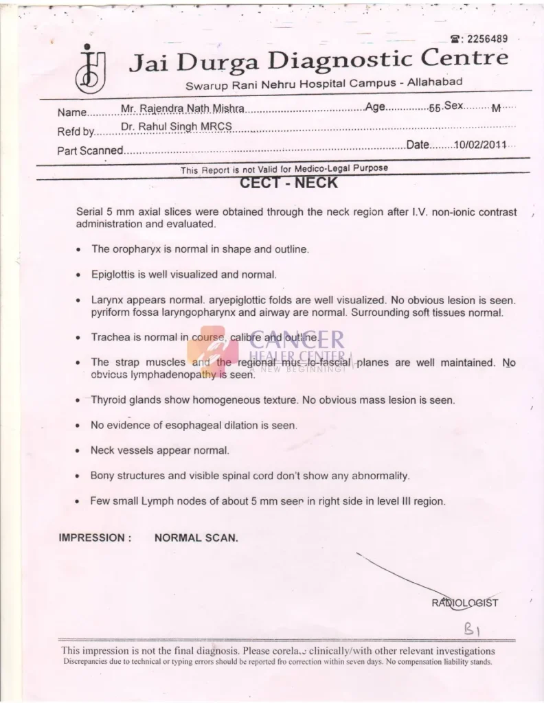
A contrast-enhanced CT scan of the neck was performed at Jai Durga Diagnostic Centre on 10 February 2011.
The report describes the major structures of the neck as normal in appearance. It identifies a few small lymph nodes measuring approximately 5 mm in the right level III region and concludes with the impression: “Normal scan.”
This conclusion reflects the radiologist’s assessment of the CT examination. It does not independently rule out a malignancy that may be identified through tissue sampling.
Cytopathology examination
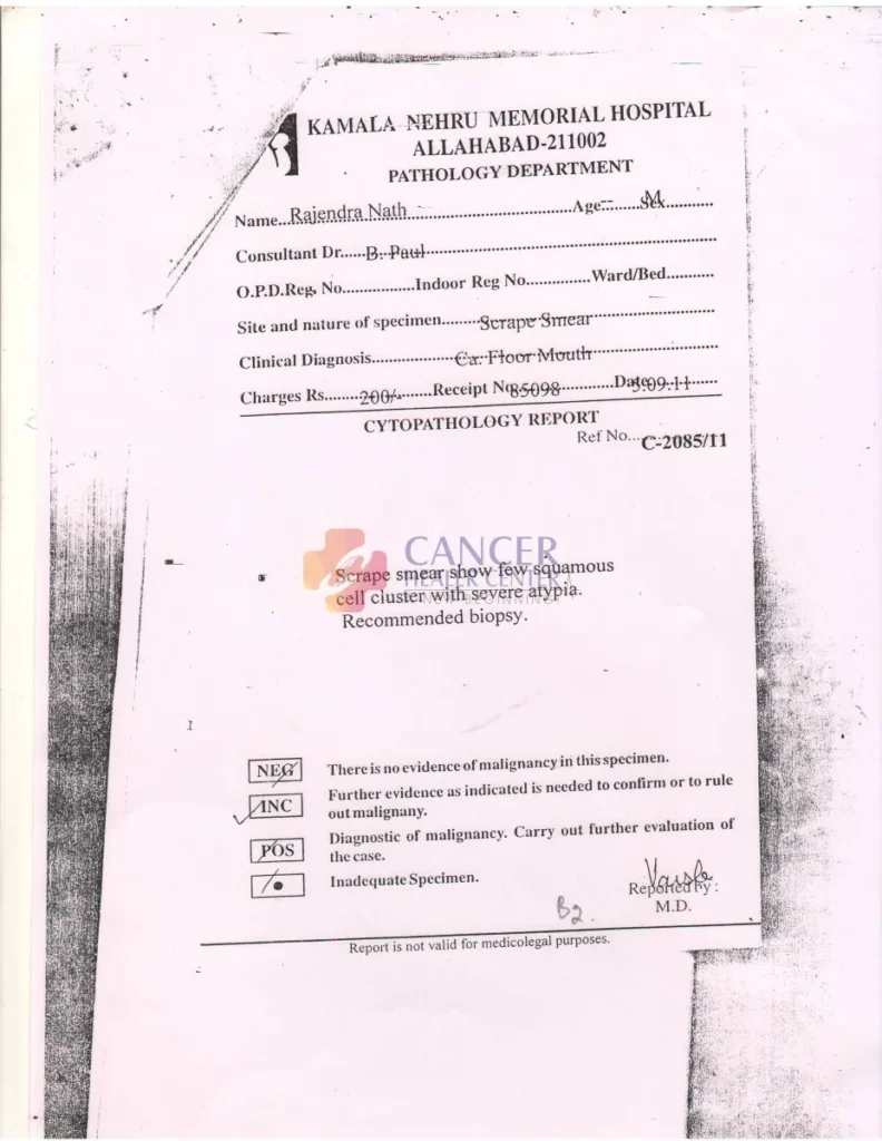
The cytopathology report from Kamala Nehru Memorial Hospital records the clinical diagnosis as cancer of the floor of the mouth.
The specimen was described as a scrape smear. The report states that the smear showed a few squamous cell clusters with severe atypia and recommends a biopsy.
Severe cellular atypia refers to significant abnormalities in the appearance of cells. The recommendation for biopsy indicates the need for further tissue evaluation.
Histopathology examination
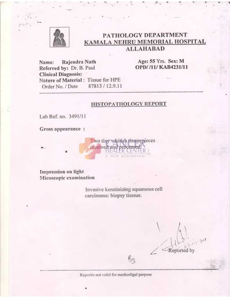
The histopathology report records an examination of biopsy tissue. The microscopic diagnosis states:
Invasive keratinizing squamous cell carcinoma: biopsy tissue.
This is the clearest diagnostic confirmation in the supplied documents.
The three reports together document an imaging assessment, a cytological finding requiring further evaluation, and a biopsy-based histopathological diagnosis.
What the Initial Neck CT Scan Showed
The CT neck examination was performed on 10 February 2011 at Jai Durga Diagnostic Centre.
The report states that serial axial images were obtained through the neck following intravenous administration of non-ionic contrast.
The radiologist documented the following findings:
- Oropharynx: Normal in shape and outline.
- Epiglottis: Well visualized and normal.
- Larynx: No obvious lesion identified.
- Aryepiglottic folds: Well visualized.
- Pyriform fossae, laryngopharynx and airway: Described as normal.
- Trachea: Normal in course, calibre and outline.
- Neck muscles and fascial planes: Well maintained.
- Thyroid gland: Homogeneous texture, with no obvious mass lesion.
- Esophagus: No dilatation identified.
- Neck vessels: Described as normal.
- Bones and visible spinal cord: No abnormality reported.
- Lymph nodes: A few small nodes, approximately 5 mm, were seen in the right level III region.
The radiologist’s impression was recorded as a normal scan.
Importantly, the report does not explicitly document a suspicious mass in the structures assessed. However, the examination’s reported findings must be interpreted alongside the subsequent or associated pathology records and the clinical examination.
Examination of the Oral and Neck Structures
The CT report describes several anatomical structures in the neck as normal in appearance.
The oropharynx, epiglottis, larynx, trachea, and surrounding soft tissues were assessed. The thyroid gland was described as homogeneous, and no obvious thyroid mass was identified.
The report also states that the neck muscles and regional musculo-fascial planes were maintained.
These findings provide information about the structures visible on the CT examination.
However, a CT scan and a biopsy answer different clinical questions. CT imaging assesses anatomical structures and may identify visible masses or other abnormalities. Histopathology examines tissue under a microscope and can identify malignant cells.
Consequently, the absence of an obvious lesion on a CT report does not invalidate a positive histopathology diagnosis.
The available records do not provide enough detail to determine the original tumour’s size, exact anatomical extent, or depth of invasion.
Lymph Nodes in the Neck
The CT neck report describes a few small lymph nodes measuring approximately 5 mm in the right level III region.
Cervical lymph nodes are part of the lymphatic system. Their assessment is important in oral cancer because squamous cell carcinoma can spread to lymph nodes in the neck.
The report does not describe these small nodes as suspicious or enlarged and concludes that the scan is normal.
Nevertheless, lymph node size alone cannot determine whether a node contains cancer. Small lymph nodes may occasionally contain malignant cells, while enlarged nodes may be caused by non-cancerous conditions.
The supplied report does not document a lymph node biopsy or provide sufficient information to establish the patient’s nodal stage.
A complete assessment would require the treating team’s clinical examination and any additional imaging, pathology, or staging investigations performed.
What the Cytopathology Report Revealed
The cytopathology report from Kamala Nehru Memorial Hospital documents a scrape smear from a site clinically identified as the floor of the mouth.
The report states:
“Scrape smear shows few squamous cell clusters with severe atypia. Recommended biopsy.”
This finding indicates that the sampled cells showed significant abnormalities and that further investigation was recommended.
Cytopathology involves the examination of individual cells or small groups of cells. It can help identify suspicious cellular changes, but its diagnostic value depends on the specimen and the findings observed.
In this case, the recommendation for biopsy was important because tissue examination could provide more definitive diagnostic information.
The associated histopathology report subsequently documents invasive keratinizing squamous cell carcinoma in the submitted biopsy tissue.
The date printed on the cytopathology report is not sufficiently clear in the supplied image and should be verified before a precise diagnostic timeline is published.
Histopathology Confirms Squamous Cell Carcinoma
The histopathology report from Kamala Nehru Memorial Hospital records the following diagnosis:
Invasive keratinizing squamous cell carcinoma: biopsy tissue.
The report describes two tiny whitish tissue pieces received and processed for microscopic examination.
The diagnosis establishes that invasive squamous cell carcinoma was identified in the submitted tissue.
What does this diagnosis mean?
Squamous cell carcinoma
This is a cancer arising from squamous epithelial cells. In the context of the clinical diagnosis recorded in the supplied report, the suspected primary site was the floor of the mouth.
Keratinizing
The term describes a microscopic feature in which tumour cells show keratin production or keratinization. This is a characteristic that pathologists may use when classifying squamous cell carcinomas.
Invasive
This indicates that malignant cells have invaded beyond the epithelial layer into underlying tissue. It distinguishes invasive carcinoma from a lesion confined to the surface epithelium.
Biopsy tissue
The diagnosis was made by examining the submitted tissue sample under a microscope.
The report confirms the presence of invasive squamous cell carcinoma in that specimen. It does not, by itself, establish the tumour’s overall size, stage, surgical margins, lymph node involvement, or presence or absence of distant metastases.
Understanding the Biopsy Findings
Histopathology plays a central role in diagnosing many cancers because it allows pathologists to examine tissue architecture and cellular characteristics.
In this case, the cytopathology report described severe atypia and recommended a biopsy. The histopathology report then documented invasive keratinizing squamous cell carcinoma.
This distinction is important.
Abnormal cells on a scrape smear may raise suspicion for malignancy and indicate the need for additional investigation. A histopathological examination can provide more definitive information about the nature of the sampled tissue.
The biopsy result in the supplied report confirms invasive squamous cell carcinoma. However, the limited description does not establish the full extent of the disease.
Additional records would be needed to determine the tumour’s dimensions, depth of invasion, grade, margin status if surgery was performed, and any involvement of nearby structures or lymph nodes.
Comparing the Imaging and Pathology Reports
The available documents describe different aspects of the patient’s evaluation.
| Investigation | Main Finding | Clinical Significance |
| CT neck, 10 February 2011 | Reported as a normal scan, with a few small right level III lymph nodes | No obvious lesion was described in the structures assessed |
| Cytopathology report | Few squamous cell clusters with severe atypia; biopsy recommended | Further tissue evaluation was advised |
| Histopathology report, 12 September 2011 | Invasive keratinizing squamous cell carcinoma identified in biopsy tissue | Confirms malignancy in the submitted specimen |
The CT and pathology findings should not be treated as contradictory without considering the clinical details and dates.
The CT report describes what was visible on the imaging examination. The histopathology report describes what was identified microscopically in the sampled tissue.
Both are valuable diagnostic tools, but neither report alone provides the complete clinical picture.
Because the exact chronology and anatomical correlation are not fully established by the supplied records, these findings should be reviewed by the treating clinician before drawing conclusions about disease progression or the relationship between the investigations.
Why Biopsy and Imaging Findings Must Be Interpreted Together
Cancer diagnosis often requires more than one type of investigation.
Imaging helps assess anatomical structures
CT scans can identify visible masses, assess surrounding anatomy, and provide information about lymph nodes and other structures within the scanned region.
However, imaging has limitations. A lesion may not be clearly visible because of its size, location, imaging characteristics, or the technical limitations of the examination.
Histopathology examines tissue directly
Biopsy involves collecting tissue for microscopic examination. This allows a pathologist to assess the cells and tissue architecture and determine whether malignancy is present in the sample.
In this case, the histopathology report explicitly confirms invasive keratinizing squamous cell carcinoma.
Clinical assessment brings the findings together
A treating clinician must interpret imaging, pathology, physical examination findings, and the patient’s medical history together.
For a suspected or confirmed floor of mouth cancer, a comprehensive assessment may include examination of the oral cavity, evaluation of the primary tumour, appropriate imaging, and assessment of cervical lymph nodes.
The exact investigations required depend on the individual case.
The key lesson from the available records is that a normal CT report should not be interpreted as excluding malignancy when tissue pathology confirms cancer.
What the Available Reports Confirm
The three reports establish the following documented findings:
- The clinical concern involved the floor of the mouth.
The cytopathology report records cancer of the floor of the mouth as the clinical diagnosis.
- The scrape smear showed significant cellular atypia.
The cytopathology report describes a few squamous cell clusters with severe atypia and recommends biopsy.
- The biopsy confirmed invasive squamous cell carcinoma.
The histopathology report identifies invasive keratinizing squamous cell carcinoma in the submitted tissue.
- The neck CT did not identify an obvious lesion in the structures described.
The radiologist reported a normal scan and documented a few small lymph nodes measuring approximately 5 mm in the right level III region.
- The available reports do not establish the complete disease stage or outcome.
There is no supplied documentation of the original tumour dimensions, full staging assessment, treatment received, subsequent imaging, or long-term follow-up.
These distinctions are essential to ensure that the published case study remains accurate and clinically responsible.
Important Clinical Information Still Required
To develop a more complete case study comparable to a detailed treatment-and-outcome article, additional clinical records would be useful.
These may include:
- The original oral examination and clinical description of the lesion.
- The date and findings of the diagnostic biopsy.
- Additional histopathology details, if available.
- Tumour size and depth of invasion.
- Clinical and pathological cancer stage.
- Assessment of cervical lymph nodes.
- Any additional imaging performed for staging.
- Details of surgery, radiotherapy, chemotherapy, immunotherapy, or other treatments received.
- Follow-up examination and imaging reports.
- Documentation of the patient’s subsequent clinical status.
Without this information, it would not be appropriate to claim that the tumour reduced in size, disappeared, responded to a specific treatment, or did not recur.
If further reports are available, they could help establish a more complete and medically verified timeline.
Conclusion
The available medical records document a case involving invasive keratinizing squamous cell carcinoma, with the clinical diagnosis on the cytopathology report referring to the floor of the mouth.
The cytopathology examination identified squamous cell clusters with severe atypia and recommended biopsy. The histopathology report subsequently documented invasive squamous cell carcinoma in the submitted tissue.
The available CT neck report, dated 10 February 2011, described the examined neck structures as normal in appearance and noted a few small lymph nodes in the right level III region.
These findings demonstrate the importance of interpreting imaging and pathology together. While imaging provides information about visible anatomical abnormalities, histopathology can establish a cancer diagnosis through microscopic tissue examination.
The available records confirm the biopsy diagnosis but do not establish the patient’s treatment course, disease stage, treatment response, or long-term outcome.
A complete clinical assessment and additional follow-up records would be necessary to determine the subsequent course of the disease.
Why Individual Assessment Matters in Oral Cancer
Oral cavity cancers can differ in their location, size, depth of invasion, pathological characteristics, and extent of spread.
For patients with floor of mouth cancer, the clinical team may need to assess the primary tumour, nearby structures, cervical lymph nodes, and other relevant areas before deciding on an appropriate treatment plan.
Important considerations include:
- The precise location and dimensions of the lesion.
- Histopathology and other relevant biopsy findings.
- Depth of invasion and involvement of nearby tissues.
- Cervical lymph node assessment.
- The patient’s overall health and medical history.
- Previous treatment and response, where applicable.
- Follow-up findings and the patient’s current clinical status.
At Cancer Healer Center, educational case studies should clearly distinguish between documented medical findings and conclusions that require additional evidence.
Any discussion of an individual patient’s diagnosis or treatment should be based on verified records and reviewed by a qualified clinician.
Patients with confirmed or suspected oral cancer should consult their treating oncology team for individualized assessment and guidance.
Medical Reports
The following records were supplied for this case study.
Report 1: CT Neck
Diagnostic centre: Jai Durga Diagnostic Centre, Allahabad
Examination date: 10 February 2011
Investigation: Contrast-enhanced CT of the neck
The report describes the major neck structures as normal in appearance and identifies a few small lymph nodes in the right level III region.
Report 2: Cytopathology Report
Hospital: Kamala Nehru Memorial Hospital, Allahabad
Specimen: Scrape smear
Clinical diagnosis: Cancer of the floor of the mouth
The report describes a few squamous cell clusters with severe atypia and recommends biopsy.
Report 3: Histopathology Report
Hospital: Kamala Nehru Memorial Hospital, Allahabad
Specimen: Tissue for histopathological examination
Report date: 12 September 2011
The microscopic diagnosis is invasive keratinizing squamous cell carcinoma in the biopsy tissue.
Publication note: Use de-identified versions of these reports in the online gallery. Remove patient names, hospital registration numbers, report identifiers, and any other personal identifiers. Confirm that authorization has been obtained for publication.
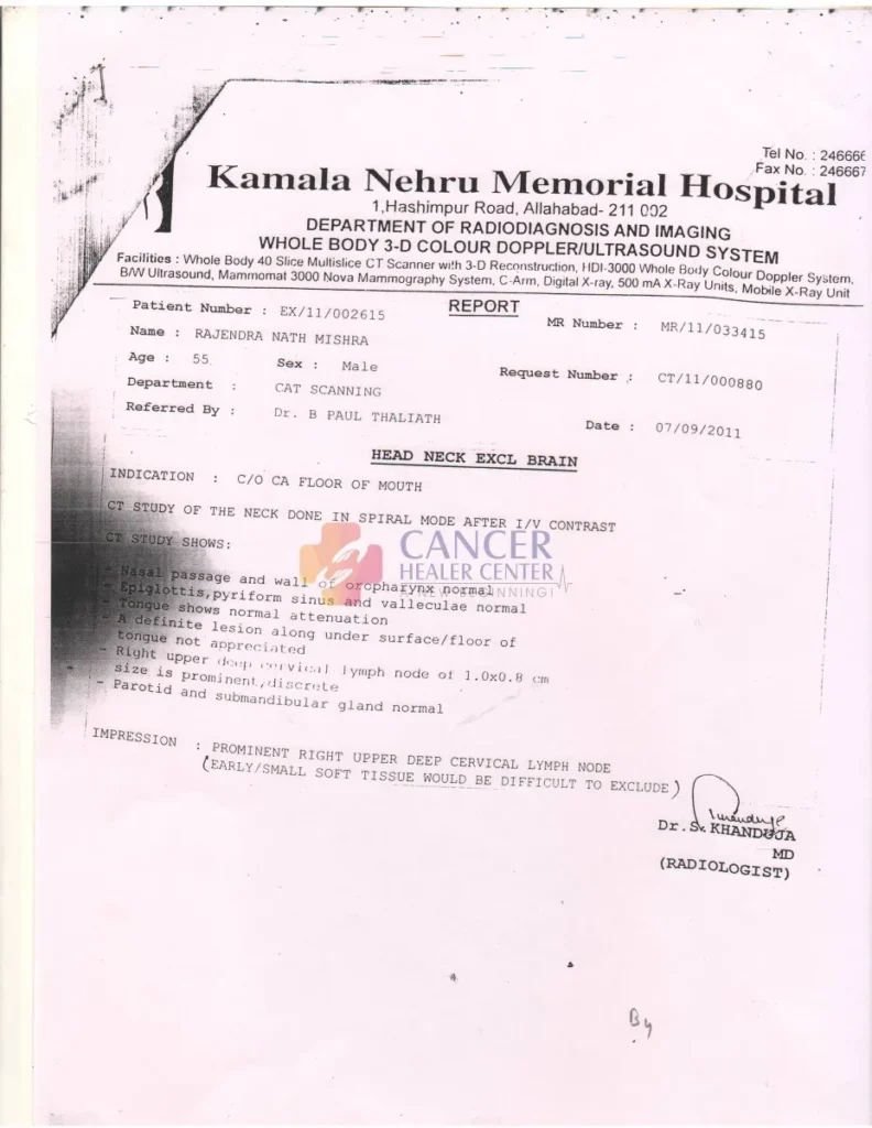
Medical disclaimer: This case study is intended for educational and informational purposes only. It is based on the available medical reports and is not a substitute for professional medical advice, diagnosis, or treatment. The findings do not establish the patient’s cancer stage, treatment response, or long-term outcome. Every patient’s condition and treatment needs are different.

Frequently Asked Questions
The histopathology report confirms invasive keratinizing squamous cell carcinoma in the submitted biopsy tissue. The associated cytopathology report records the clinical diagnosis as cancer of the floor of the mouth.
It is a cancer arising from squamous cells in the lining of the oral cavity. The floor of the mouth is the area beneath the tongue. The diagnosis and extent of disease must be established through appropriate clinical examination and investigations.
The scrape smear showed a few squamous cell clusters with severe atypia. The report recommended a biopsy for further evaluation.
The report identified invasive keratinizing squamous cell carcinoma in the submitted biopsy tissue.
The radiologist did not identify an obvious lesion in the neck structures described in the report. However, imaging and histopathology assess different aspects of a suspected cancer. A normal-appearing CT scan does not exclude a malignancy confirmed by biopsy.
The CT report described a few small lymph nodes measuring approximately 5 mm in the right level III region. The report did not characterize them as suspicious. Their clinical significance cannot be determined from size alone.
No. The histopathology report confirms cancer in the sampled tissue but does not provide enough information to determine the complete stage. Staging requires additional clinical, pathological, and imaging information as appropriate.
No. The supplied documents do not include a treatment history or post-treatment assessment. They establish diagnostic findings but do not demonstrate a treatment response.
The original clinical examination, complete pathology findings, staging investigations, treatment records, and subsequent follow-up reports would help establish the patient’s clinical timeline and outcome.
They should consult a qualified oncology team for assessment of the primary tumour, appropriate staging investigations, and discussion of treatment options. Treatment decisions should be based on the individual’s diagnosis, stage, health, and clinical circumstances.