
A Strong Response Documented Across Follow-Up Scans
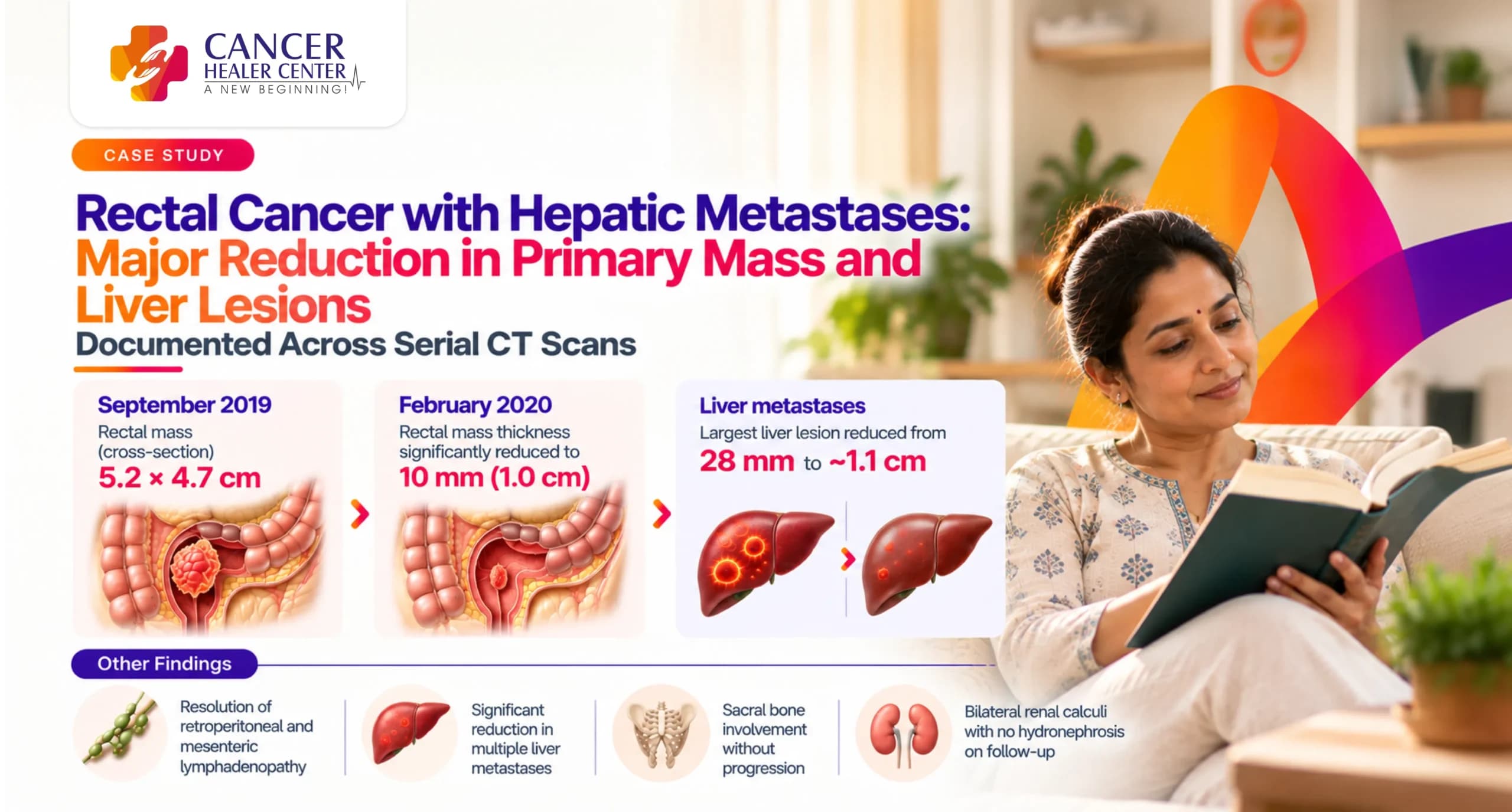
Metastatic rectal carcinoma with liver involvement represents an advanced oncological challenge requiring continuous longitudinal monitoring. This case details the clinical journey of Mrs. Sarita Pal, a 38-year-old female diagnosed with rectal cancer with hepatic metastases. Serial Contrast-Enhanced Computed Tomography (CECT) scans performed between September 2019 and February 2020 document a major treatment response, featuring dramatic shrinkage of both the primary rectal mass and metastatic liver lesions alongside resolution of regional lymphadenopathy.
Mrs. Sarita Pal’s Case at a Glance
- Patient Name: Mrs. Sarita Pal
- Age / Sex: 38 Years / Female
- Diagnosis: Metastatic Rectal Carcinoma with Liver Metastases (Ca Rectum with Liver Mets)
- Primary Findings at Baseline (19/09/2019): Circumferential rectal mass (5.5 cm length, 5.2 x 4.7 cm cross-section) with loss of fat planes with uterus/cervix; sacral cortical erosion; infrarenal paraaortic (27 x 24 mm) and presacral (13–15 mm) lymph nodes; multiple liver metastases up to 28 mm.
- Key Scans Evaluated: CT Whole Abdomen Contrast (19/09/2019) — Atulaya Healthcare; CECT Chest & Abdomen (24/02/2020) — P.G.I.M.E.R., Chandigarh.
- Primary Outcome: Reduction of primary rectal mass thickness to 10 mm (from 5.2 x 4.7 cm cross-section); dramatic reduction in liver lesion size from 28 mm down to ~1.1 cm; complete resolution of retroperitoneal/mesenteric lymphadenopathy.
Mrs. Sarita Pal’s Treatment Journey
Mrs. Sarita Pal presented in September 2019 with abdominal pain and rectal bleeding. Baseline contrast CT imaging confirmed a locally advanced rectal mass with extensive sacral invasion, retroperitoneal lymphadenopathy, and multiple bilateral liver metastases. In September 2019, she initiated treatment under Cancer Healer Center with Dr. Tarang Krishna. A follow-up 128-slice CECT scan of the chest and abdomen was conducted at P.G.I.M.E.R., Chandigarh in February 2020 to evaluate treatment response.
What the September 2019 Baseline CT Scan Showed
A CT Whole Abdomen Contrast scan was performed on 19/09/2019 at Atulaya Healthcare, Chandigarh.
Primary Rectal Mass & Pelvic Involvement in September 2019
- Primary Mass Dimensions: Lobulated heterogenous nearly circumferential mass of approx 5.5 cm length (approx 8.5 cm from anal verge) with right-sided exogenous tumor bulk measuring 5.2 x 4.7 cm. Adjacent satellite nodules of 8–10 mm noted.
- Infiltration & Bone Erosion: Loss of fat plane with lower uterine/cervix region. Sacrum 3, 4, and 5 vertebrae showed focal rarefied appearance with mild cortical erosion and adjacent soft tissue (bony metastasis). Presacral lymphnodes/nodules of 13–15 mm observed.
Hepatic Metastases & Lymphadenopathy in September 2019
- Liver Metastases: Multiple hypoenhancing soft tissue density lesions in both lobes of the liver, the largest measuring 28 mm, representing metastases.
- Retroperitoneal Lymph Nodes: Infrarenal left paraaortic soft tissue density mildly enhancing lymph node measuring 27 x 24 mm.
- Kidneys: Bilateral renal calculi (up to 6.0 mm in right kidney, 5.0 mm in left kidney) with prominent right ureter.
What the February 2020 Follow-Up CT Scan Confirmed
A follow-up CECT of the chest and abdomen was performed on 24/02/2020 at P.G.I.M.E.R., Chandigarh using a 128-slice scanner.
Major Shrinkage of Primary Rectal Tumor by February 2020
- Primary Mass Regressive Changes: Asymmetrical mural thickening with maximum thickness of only 10 mm (1.0 cm) in the rectum (5 cm segment, 2.5 cm from anal verge), representing a dramatic decrease from the initial 5.2 x 4.7 cm cross-sectional mass.
- Surrounding Tissue Planes: Ill-defined fat planes preserved with mesorectal fascia and uterus without aggressive soft tissue invasion.
Significant Regression of Liver Metastases
- Liver Lesions: Multiple well-defined hypodense lesions remained studded in bilateral liver lobes, but the largest lesion measured only ~1.1 cm x 1.0 cm in segment II (reduced from 28 mm at baseline). Normal craniocaudal span (14.2 cm).
Resolution of Lymphadenopathy & Distant Spread
- Lymph Nodes: No significant retroperitoneal or mesenteric adenopathy seen (complete clearance of the 27 x 24 mm paraaortic node). Subcentimetric pretaracheal/subcarinal nodes in chest.
- Chest & Abdomen: Both lungs normal with no pleural/pericardial effusion. Spleen, pancreas, adrenals, gallbladder, and urinary bladder normal.
The Response Documented Over Time
| Target Region / Feature | September 2019 Baseline CT | February 2020 Follow-Up CT | Overall Clinical Response |
| Primary Rectal Mass | 5.5 cm length mass, 5.2 x 4.7 cm tumor bulk | Asymmetrical thickening (Max 10 mm thickness) | Significant Tumor Shrinkage |
| Hepatic Metastases | Multiple bilateral lesions, largest 28 mm | Multiple well-defined lesions, largest ~1.1 x 1.0 cm | Major Volumetric Reduction |
| Paraaortic Lymph Node | Infrarenal left paraaortic node 27 x 24 mm | No significant retroperitoneal adenopathy | Completely Resolved |
| Presacral Nodes / Infiltration | Presacral nodes 13–15 mm with sacral cortical erosion | Ill-defined fat planes preserved with mesorectal fascia | Marked Infiltration Regression |
| Chest / Lung Parenchyma | Not evaluated | Normal lung volume & broncho-vascular pattern | No Distant Thoracic Spread |
What the Two Reports Confirm
- 1. Marked Local Primary Contraction: The large bulky rectal mass measuring 5.2 x 4.7 cm in cross-section shrank significantly down to a residual asymmetrical mural thickening of max 10 mm.
- 2. Shrinkage of Liver Metastases: The maximal liver lesion diameter reduced by over 60% (from 28 mm down to 11 mm) with well-defined contours.
- 3. Resolution of Retroperitoneal Disease: The large 27 x 24 mm infrarenal paraaortic lymph node re-cleared completely on follow-up imaging.
Understanding the CT Scan Findings
- Mural Thickening vs Mass Effect: Transitioning from a solid exophytic mass extending into uterine fat planes to a focal 10 mm mural wall thickening confirms disease regression and relief of pelvic mass effect.
- Systemic Metastatic Control: Concomitant reduction in hepatic tumor burden reconfirms effective systemic disease control.
Patient Testimonial
In a handwritten testimonial letter, Mrs. Sarita Pal expressed her satisfaction with her treatment at Cancer Healer Center:
“मेरा नाम सरिता पाल w/o विजय कुमार पाल। मुझे रेक्टम में कैंसर की परेशानी थी। मेरा इलाज डॉ कृष्णा कैंसर सेटर से 8 सितम्बर 2019 से चल रहा है। मुझे पहले से काफी राहत है और मेरे सीटी स्कैन में भी बीमारी में पहले से कुछ सुधार दिखा है। — सरिता पाल”
Conclusion
This serial CECT evaluation confirms a strong objective treatment response in a patient with advanced metastatic rectal cancer. The major contraction of the primary pelvic mass combined with significant regression of liver metastases and node clearance demonstrates clear therapeutic efficacy.
Every Rectal Cancer Case Requires Individual Assessment
Disclaimer: Every cancer case involves unique diagnostic, histological, and anatomical factors. Treatment plans and radiological outcomes vary per individual. Clinical correlation and medical consultation remain essential for evaluating patient treatment protocols.
Medical Reports
Below are the official CT diagnostic reports and testimonial documentation for Mrs. Sarita Pal:
- CT Whole Abdomen Contrast (19/09/2019) — Page 1 (Atulaya Healthcare)
- CT Whole Abdomen Contrast (19/09/2019) — Page 2 (Atulaya Healthcare)
- CECT Chest & Abdomen (24/02/2020) — Follow-Up Report (P.G.I.M.E.R., Chandigarh)
- Patient Testimonial Letter (Handwritten Feedback)
Medical Reports
Frequently Asked Questions
Mrs. Sarita Pal was diagnosed with metastatic rectal carcinoma (Ca rectum) with bilateral liver metastases, sacral bone involvement, and retroperitoneal lymphadenopathy.
In September 2019, the CT scan showed a 5.5 cm long circumferential rectal mass with exogenous tumor bulk measuring 5.2 x 4.7 cm causing loss of fat planes with the uterine cervix.
The follow-up CECT scan at P.G.I.M.E.R. Chandigarh showed that the primary mass shrank to a residual asymmetrical mural thickening of max 10 mm, the largest liver lesion shrank from 28 mm down to 1.1 cm, and retroperitoneal lymph nodes completely cleared.
The previously noted 27 x 24 mm infrarenal left paraaortic lymph node and presacral nodes resolved completely, with no significant adenopathy reported in February 2020.