
A Strong Response Documented Across Serial Scans
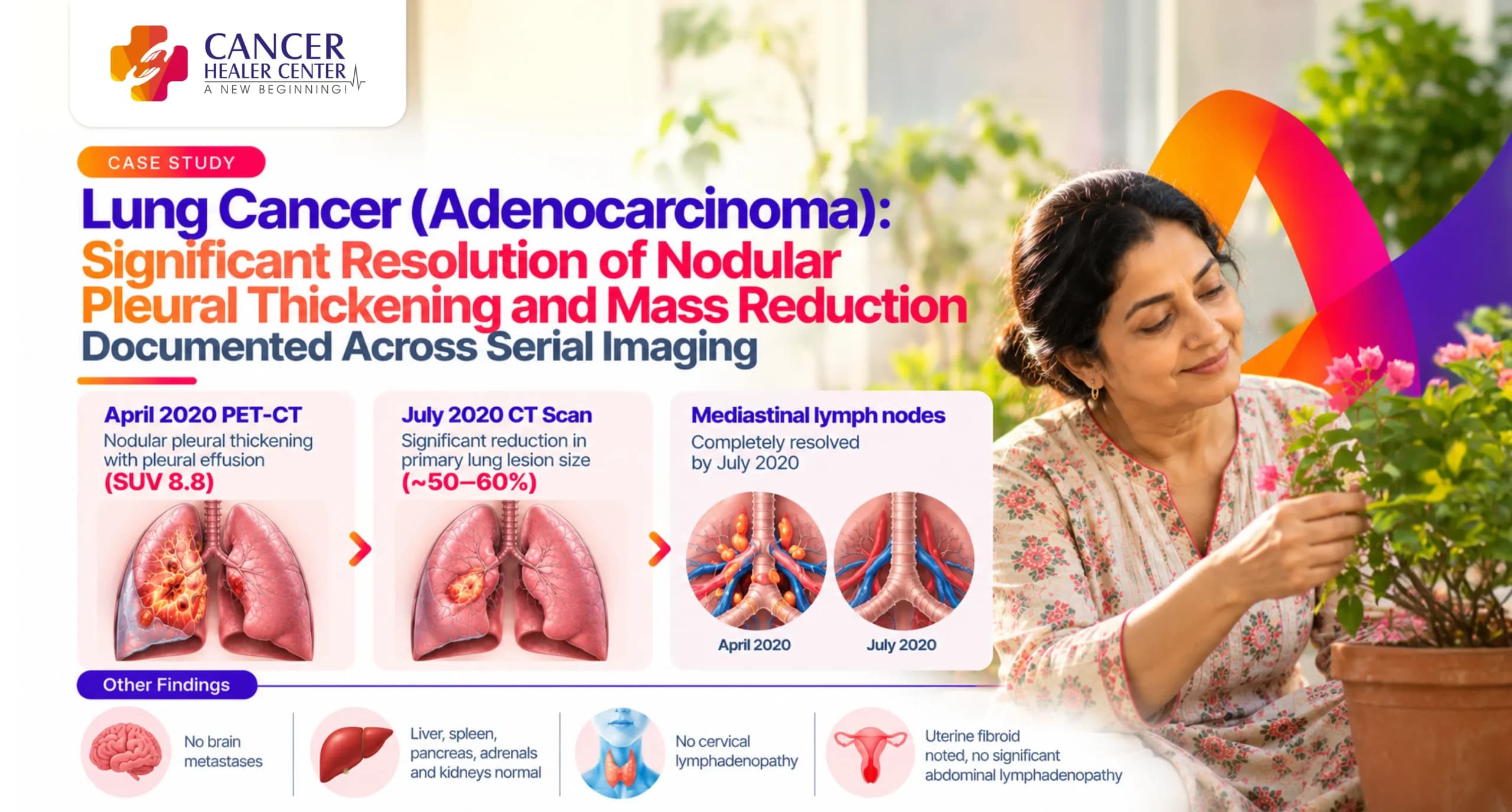
Lung adenocarcinoma with pleural and nodal involvement poses significant management challenges requiring ongoing serial imaging monitoring. This case details the clinical progress of Mrs. Rizwana Begum, a 55-year-old female diagnosed with lung adenocarcinoma, who demonstrated a major objective response to therapy between April 2020 and July 2020. Serial 18F-FDG PET-CT and contrast-enhanced chest CT scans document significant resolution of nodular pleural thickening, reduction in pleural effusion, complete clearance of mediastinal lymphadenopathy, and marked regression of the primary parenchymal lesion.
Mrs. Rizwana Begum’s Case at a Glance
- Patient Name: Mrs. Rizwana Begum
- Age / Sex: 55 Years / Female
- Diagnosis: Carcinoma Lung / Adenocarcinoma of the Lung with Mediastinal Lymph Node Metastosis
- Primary Findings at April 2020 PET-CT: Passive collapse of right middle and lower lobes with abnormal FDG uptake (max SUV 8.6); right moderate lobulated pleural effusion with diffuse nodular pleural thickening (max SUV 8.8); subcentimetric mediastinal nodes (max SUV 3).
- Key Scans Evaluated: Whole Body 18F-FDG PET-CT Scan (06/04/2020) — Vijaya Diagnostic Centre; Contrast-Enhanced CT Thorax (23/07/2020) — Advanced Diagnostic Centre.
- Primary Outcome: Substantial reduction (50–60%) in primary lung lesion size; complete resolution of mediastinal lymphadenopathy; marked decrease in right pleural effusion and nodular pleural thickening.
Mrs. Rizwana Begum’s Treatment Journey
Mrs. Rizwana Begum was diagnosed with lung carcinoma and mediastinal lymph node metastasis in 2018 and initially received conventional chemotherapy. Seeking integrative options, her family initiated care under Cancer Healer Center with Dr. Tarang Krishna in April 2020. Standardized baseline PET-CT imaging was performed in April 2020, followed by a high-resolution contrast-enhanced CT scan of the thorax in July 2020 to assess therapeutic response.
What the April 2020 PET-CT Scan Showed
A whole-body 18F-FDG PET-CT scan was performed on 06/04/2020 at Vijaya Diagnostic Centre, Hyderabad.
Brain, Neck, and Abdominal Findings in April 2020
- Brain: Normal parenchymal attenuation without evidence of space-occupying lesions or brain metastases.
- Neck: Normal aerodigestive tract, salivary glands, and thyroid gland. No cervical lymphadenopathy.
- Abdomen & Pelvis: Normal liver contour with a small non-FDG-avid hypodensity. Normal gallbladder, pancreas, spleen, adrenals, kidneys, and gastrointestinal tract. Uterine fibroid noted. No ascites or significant abdominal lymphadenopathy.
Chest and Pleural Findings in April 2020
- Pleural Involvement: Right moderate lobulated pleural effusion with diffuse nodular pleural thickening showing increased tracer uptake (max SUV 8.8).
- Parenchymal Mass & Collapse: Passive collapse of the right middle and lower lobes with an area of abnormal FDG uptake within the collapsed lower lobe measuring ~2 x 1.3 cm (max SUV 8.6). Atelectatic changes in the left lingular segment.
- Mediastinal Nodes: Subcentimetric mediastinal lymph nodes displaying mild FDG uptake (max SUV 3).
Impression of the April 2020 PET-CT Scan
- 1. Passive collapse of right middle/lower lobe with abnormal FDG uptake — concern for recurrence.
- 2. Right moderate lobulated pleural effusion with diffuse nodular pleural thickening showing increased FDG uptake.
- 3. Subcentimetric mediastinal nodes with mild uptake.
- Comparison: In comparison to 03/10/2019 PET-CT, showed increased FDG uptake in the right lower lobe lesion, new right pleural effusion/thickening, and minimal increase in mediastinal node uptake.
What the July 2020 Follow-Up CT Scan Confirmed
A follow-up Multislice Contrast-Enhanced CT (CECT) of the thorax was conducted on 23/07/2020.
Mediastinum and Airway Evaluation in July 2020
- Airways: Trachea and major bronchi were normal in caliber.
- Mediastinal Nodes: No significant mediastinal lymphadenopathy was observed, confirming complete resolution of earlier nodal activity.
- Vascular Structures: Mediastinal vascular structures appeared within normal limits.
Significant Pleural and Mass Resolution by July 2020
- Right Upper Lobe: Subsegmental collapse of the right upper lobe with fissural-based thickening and a fissural-based nodule measuring 12 x 11 mm. Associated mild pleural thickening in the anterior segment.
- Right Lower Lobe: Subsegmental collapse with focal areas of cicatricial fibro-bronchiectatic changes.
- Pleural Effusion: Reduced to only mild right pleural effusion.
Impression of the July 2020 Contrast CT Scan
- • Significant resolution of nodular pleural thickening and right pleural effusion.
- • Marked reduction in the size of the heterogenous mass lesion as compared to the April 2020 PET-CT scan.
- • Overall features indicative of Good Treatment Response.
The Response Documented Over Time
| Target Region / Feature | April 2020 PET-CT Scan | July 2020 CECT Thorax Scan | Overall Clinical Outcome |
| Primary Lung Mass | Passive collapse with active lesion (20 x 13 mm, SUV 8.6) | 12 x 11 mm fissural nodule with partial collapse | 50–60% Size Reduction |
| Mediastinal Lymph Nodes | Subcentimetric nodes with FDG uptake (SUV 3) | No significant lymphadenopathy | Completely Resolved |
| Pleural Effusion | Moderate lobulated right effusion | Mild right effusion | Marked Reduction |
| Pleural Thickening | Diffuse nodular pleural thickening (SUV 8.8) | Mild residual pleural thickening | Significant Resolution |
| Distant Metastases | None (Brain, Abdomen clear) | None identified | Sustained Systemic Control |
What the Serial Reports Confirm
The head-to-head objective comparison between the April 2020 baseline PET-CT and July 2020 follow-up CECT confirms:
- 1. Regression of Primary Tumor & Nodes: Clear volumetric contraction of the pulmonary lesion alongside complete clearance of suspicious mediastinal lymph nodes.
- 2. Control of Pleural Disease: Dramatic reduction in both pleural fluid volume and nodular thickening along the right pleural surfaces.
Understanding the PET-CT and CT Scan Findings
- FDG Metabolic Activity vs. Structural Size: The high metabolic activity (SUV max up to 8.8) noted in April 2020 reflected active malignancy. Structural regression by July 2020 confirms functional tumor control and tissue shrinkage.
- Resolution of Effusion: The regression from moderate lobulated effusion to minimal fluid accumulation correlates directly with reduced pleural tumor burden.
Patient Testimonial
In a written letter dated 29th July 2020, Mohammad Javeed Ali (son of Mrs. Rizwana Begum) shared their treatment experience:
“My mother was diagnosed with CA Lung with mediastinal lymph node metastasis since 2018… We came to know about Dr. Tarang Krishna’s Cancer Healer Center & started taking treatment since April 2020 at Hyderabad center. Taking the medicines regularly since then, we had CT-Scan chest report done in July 2020 and we could see 50-60% reduction in the lung lesion and lymphnode are resolved completely. There is a reduction in the pleural effusion and thickening in right lung. I am thankful to Dr. Tarang sir & his team of doctors…”
Conclusion
The objective diagnostic imaging timeline confirms a significant positive response to treatment in a patient with recurrent lung adenocarcinoma. The complete clearance of mediastinal lymphadenopathy combined with a major contraction in primary tumor burden and pleural involvement highlights substantial clinical recovery and structural disease control.
Every Lung Cancer Case Requires Individual Assessment
Disclaimer: Every cancer case involves unique diagnostic, histological, and anatomical factors. Treatment plans and radiological outcomes vary per individual. Clinical correlation and medical consultation remain essential for evaluating patient treatment protocols.
Medical Reports
Below is the record of diagnostic reports and documentation for Mrs. Rizwana Begum:
- 1. PET-CT Scan Report (06/04/2020) — Page 1 (Vijaya Diagnostic Centre)
- 2. PET-CT Scan Report (06/04/2020) — Page 2 (Vijaya Diagnostic Centre)
- 3. PET-CT Scan Report (06/04/2020) — Page 3 (Vijaya Diagnostic Centre)
- 4. CECT Chest Report (23/07/2020) — Findings Page (Advanced Diagnostic Centre)
- 5. CECT Chest Report (23/07/2020) — Impression Page (Advanced Diagnostic Centre)
- 6. Handwritten Patient Testimonial Letter (29/07/2020)
Frequently Asked Questions
Mrs. Rizwana Begum was diagnosed with Adenocarcinoma of the Lung (CA Lung) with mediastinal lymph node metastasis and right pleural involvement.
The scan showed an active right lower lobe mass/collapse (~2 x 1.3 cm, max SUV 8.6), moderate lobulated right pleural effusion with diffuse nodular pleural thickening (max SUV 8.8), and mild FDG-avid subcentimetric mediastinal lymph nodes.
The CECT scan revealed complete resolution of mediastinal lymphadenopathy, significant regression of nodular pleural thickening, reduction of right pleural effusion to mild, and a 50–60% reduction in primary lesion size.
No. Whole-body imaging confirmed that the brain, liver, kidneys, and abdominal organs were free of metastatic disease.