


A review of two abdominal CT reports documenting postoperative findings following gallbladder cancer surgery, with follow-up imaging in August 2011 and March 2012.
This case study examines the imaging findings of a patient who underwent surgery for gallbladder carcinoma, including cholecystectomy and focal hepatic resection. The available CT reports document the postoperative appearance of the gallbladder fossa, changes in liver appearance, and additional findings involving the kidneys and spine.
The March 2012 report specifically states that no residual enhancing mass was identified in the gallbladder fossa. The findings provide a documented picture of the patient’s abdominal status at the time of the follow-up scan.
Clinical note: This case study is based on the two available CT reports. Histopathology, staging records, treatment details, and subsequent follow-up are not available for review. The imaging findings should therefore be interpreted within these limitations.
Table of Contents
- A Follow-Up Imaging Review After Gallbladder Cancer Surgery
- The Patient’s Case at a Glance
- The Patient’s Medical Journey
- What the August 2011 CT Scan Showed
- Liver Findings in August 2011
- Other Abdominal Findings in August 2011
- What the March 2012 CT Scan Showed
- No Residual Enhancing Mass in the Gallbladder Fossa
- Liver Findings on Follow-Up Imaging
- Bilateral Kidney Stones
- Other Findings: Abdomen, Pelvis and Spine
- Comparing the Two CT Reports
- What the Imaging Findings Tell Us
- Conclusion
- Why Individual Assessment Matters in Gallbladder Cancer
- Medical Reports
- Frequently Asked Questions
- A Follow-Up Imaging Review After Gallbladder Cancer Surgery
Gallbladder cancer requires careful evaluation and follow-up based on the disease stage, surgical findings, pathology results, and the patient’s overall clinical condition.
In this case, the available medical records describe a patient who underwent cholecystectomy and focal hepatic resection for gallbladder carcinoma. Two CT reports provide information about the postoperative abdominal findings at different points in time.
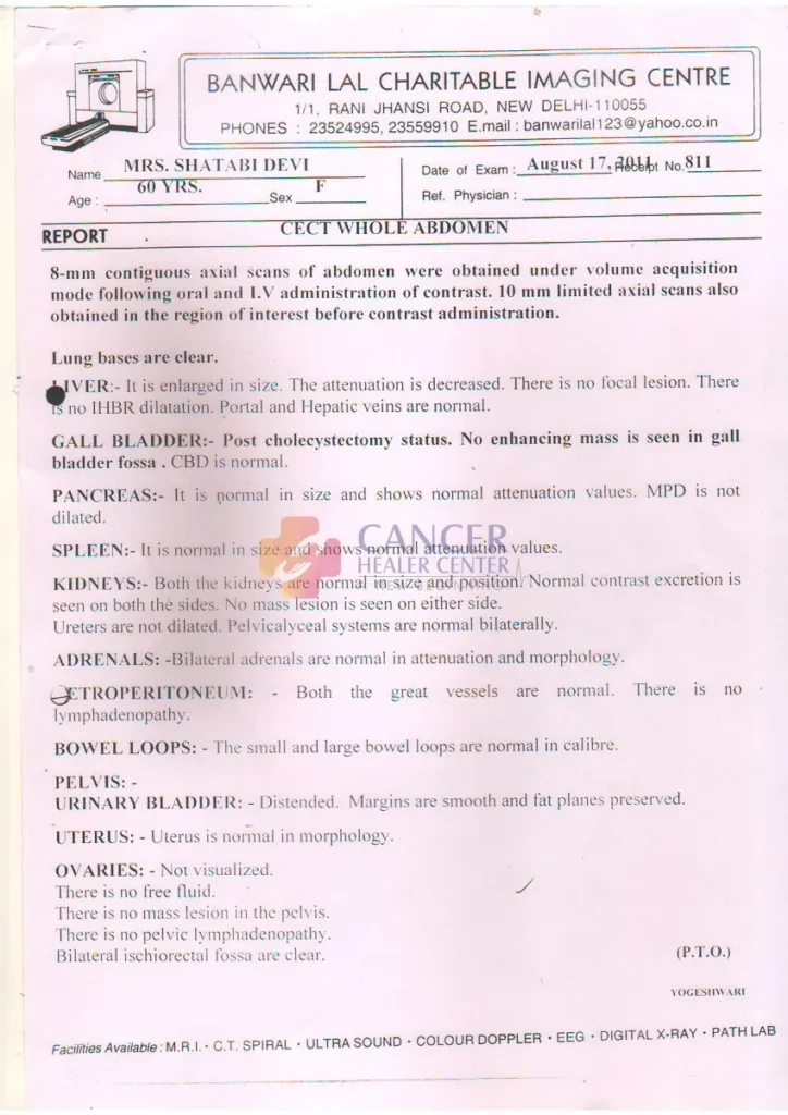
The first report, dated August 17, 2011, describes an enlarged liver with decreased attenuation, consistent with a fatty change in the liver. It also documents the postoperative absence of the gallbladder and no focal liver lesion.
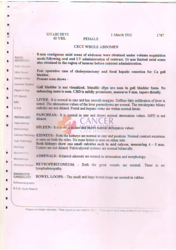
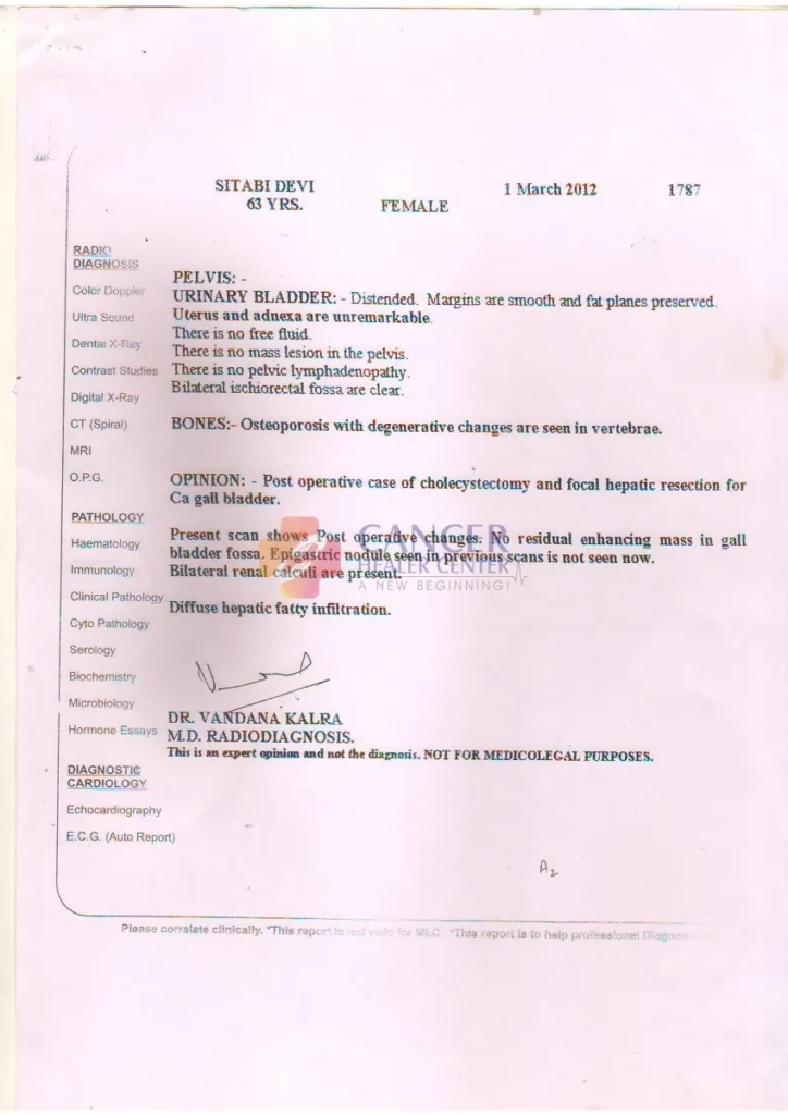
The second report, dated March 1, 2012, explicitly identifies the patient as a postoperative case of gallbladder cancer surgery. It reports no residual enhancing mass in the gallbladder fossa and notes that previously described epigastric nodular soft tissue was no longer visible.
The later report also documents diffuse fatty infiltration of the liver and small stones in both kidneys.
Together, these reports provide a record of postoperative imaging findings. They do not, by themselves, establish long-term cancer status or the effect of any particular treatment.
- The Patient’s Case at a Glance
| Clinical Detail | Information Documented |
| Primary diagnosis | Gallbladder carcinoma |
| Surgical history | Cholecystectomy and focal hepatic resection |
| First available CT report | August 17, 2011 |
| Second available CT report | March 1, 2012 |
| Imaging modality | Contrast-enhanced CT of the whole abdomen |
| Gallbladder fossa | No enhancing mass reported in August 2011; no residual enhancing mass reported in March 2012 |
| Liver | Decreased attenuation in August 2011; diffuse fatty infiltration described in March 2012 |
| Lymph nodes | No retroperitoneal lymphadenopathy reported |
| Additional findings | Bilateral renal calculi on the March 2012 scan |
| Treatment details | Not available in the supplied reports |
| Long-term outcome | Not documented in the available records |
Note: The patient’s name and age differ between the two reports. Demographic details should be verified against the original medical record before publication.
- The Patient’s Medical Journey
The available documentation describes the following sequence of events:
Gallbladder cancer diagnosis and surgery
The March 2012 report identifies the patient as having undergone cholecystectomy and focal hepatic resection for gallbladder carcinoma.
These procedures involve removal of the gallbladder and surgical removal of a portion of liver tissue, respectively. The precise extent of surgery and the original disease stage cannot be determined from the supplied CT reports alone.
August 17, 2011: First available abdominal CT report
The CT examination documented postoperative changes, an enlarged liver with decreased attenuation, and no focal liver lesion. No enhancing mass was identified in the gallbladder fossa.
March 1, 2012: Follow-up abdominal CT report
The later report described postoperative changes, no residual enhancing mass in the gallbladder fossa, and no previously described epigastric nodular soft tissue visible on the current scan.
It also documented diffuse fatty infiltration of the liver and bilateral renal calculi.
These dates establish the sequence of the available imaging records. The reports do not provide a complete account of the patient’s treatment between scans.
- What the August 2011 CT Scan Showed
The first available report was issued by Banwari Lal Charitable Imaging Centre on August 17, 2011.
The examination involved contrast-enhanced CT imaging of the whole abdomen.
The report described the following findings:
- Lung bases: Clear.
- Liver: Enlarged, with decreased attenuation and no focal lesion.
- Biliary system: No intrahepatic biliary radicle dilatation; portal and hepatic veins were described as normal.
- Gallbladder fossa: Post-cholecystectomy status, with no enhancing mass identified.
- Common bile duct: Reported as normal.
- Pancreas: Normal in size and attenuation, without pancreatic duct dilatation.
- Spleen: Normal in size and attenuation.
- Kidneys: Normal in size and position, without a mass lesion.
- Adrenal glands: Normal in appearance.
- Retroperitoneum: No lymphadenopathy reported.
- Bowel loops: Normal in calibre.
- Pelvis: No pelvic mass, free fluid, or pelvic lymphadenopathy reported.
The report therefore documented postoperative findings without identifying an enhancing mass in the gallbladder fossa at that examination.
- Liver Findings in August 2011
The August 2011 report described an enlarged liver with decreased attenuation.
Decreased attenuation on CT can be associated with fatty infiltration of the liver. However, imaging findings must be interpreted alongside the patient’s clinical history and other investigations.
The report did not identify a focal liver lesion. It also described the portal and hepatic veins as normal and noted no intrahepatic biliary dilatation.
These observations are relevant to the assessment of the liver and biliary system following surgery for gallbladder cancer.
The report does not establish whether the liver changes were related to the cancer, previous treatment, a separate liver condition, or another cause.
- Other Abdominal Findings in August 2011
The first CT report documented generally unremarkable findings in several abdominal and pelvic structures.
The pancreas, spleen, kidneys, and adrenal glands were described as normal. The bowel loops were also reported as normal in calibre.
No retroperitoneal lymphadenopathy was identified. The pelvic assessment did not show free fluid, a pelvic mass, or pelvic lymphadenopathy.
These findings describe the structures examined on that particular scan. They should not be interpreted as proof that cancer was absent from every part of the body.
- What the March 2012 CT Scan Showed
The second report, dated March 1, 2012, provides additional information about the patient’s postoperative status.
It explicitly describes the patient as a postoperative case of cholecystectomy and focal hepatic resection for gallbladder carcinoma.
The report records the following findings:
- The gallbladder was not visualized, with metallic clips seen in the gallbladder fossa.
- No enhancing mass was identified in the gallbladder fossa.
- The common bile duct was mildly prominent, measuring approximately 9 mm and tapering distally.
- The liver was normal in size with smooth margins.
- Diffuse fatty infiltration of the liver was noted.
- The intrahepatic biliary radicles were not dilated.
- The pancreas, spleen, and adrenal glands were described as normal.
- No retroperitoneal lymphadenopathy was identified.
- Small renal calculi measuring approximately 4–5 mm were present in both kidneys.
- No pelvic mass, free fluid, or pelvic lymphadenopathy was identified.
- Osteoporosis with degenerative changes was noted in the vertebrae.
The report’s principal postoperative observation was the absence of a residual enhancing mass in the gallbladder fossa.
- No Residual Enhancing Mass in the Gallbladder Fossa
One of the key observations in the March 2012 CT report concerns the gallbladder fossa, the area where the gallbladder was previously located.
The radiologist documented postoperative changes, including metallic clips, and stated that no residual enhancing mass was seen.
The report also noted that epigastric nodular soft tissue described on previous scans was not visible on the current examination.
These findings are relevant when reviewing postoperative imaging following gallbladder cancer surgery.
However, the absence of an enhancing mass on CT does not independently establish that all cancer cells have been eliminated. Microscopic disease and abnormalities below the detection limits of imaging may not be visible.
A complete assessment of cancer status requires consideration of the original pathology, surgical margins, disease stage, clinical examination, and any subsequent investigations.
- Liver Findings on Follow-Up Imaging
The March 2012 report described the liver as normal in size, with smooth margins and diffuse fatty infiltration.
It also stated that the liver parenchyma had normal attenuation values, despite noting diffuse fatty infiltration. These descriptions should be reviewed against the original images if a detailed radiological interpretation is required.
The intrahepatic biliary radicles were not dilated, and the portal and hepatic veins were reported to be within normal limits.
The common bile duct was mildly prominent, measuring approximately 9 mm and tapering distally.
The clinical significance of this measurement depends on the patient’s surgical history, age, symptoms, laboratory results, and other imaging findings. It should not be interpreted in isolation.
- Bilateral Kidney Stones
The March 2012 CT report identified small calculi in both kidneys, measuring approximately 4–5 mm.
Renal calculi are mineral deposits that form within the kidneys. Depending on their location and whether they obstruct urine flow, they may cause no symptoms or may be associated with pain, blood in the urine, or urinary complications.
The report stated that the ureters were not dilated and the pelvicalyceal systems were normal bilaterally.
These findings do not suggest urinary tract dilatation on the reported scan, but the kidney stones should be considered separately from the patient’s gallbladder cancer history.
Any management decision would depend on symptoms, kidney function, stone location, and a clinician’s assessment.
- Other Findings: Abdomen, Pelvis and Spine
The March 2012 report also documented several additional findings.
Pancreas and spleen
Both organs were described as normal in size and attenuation, with no pancreatic duct dilatation.
Adrenal glands
The adrenal glands were reported to be normal in attenuation and morphology.
Retroperitoneum
The great vessels were described as normal, and no retroperitoneal lymphadenopathy was identified.
Bowel and pelvis
The bowel loops were normal in calibre. The uterus and adnexa were described as unremarkable. No pelvic mass, free fluid, or pelvic lymphadenopathy was reported.
Bones
Osteoporosis with degenerative changes in the vertebrae was noted.
These observations provide additional context about the patient’s abdominal and pelvic status at the time of the March 2012 examination. They do not replace a comprehensive clinical assessment.
- Comparing the Two CT Reports
Reviewing the two reports together helps establish what was documented at each examination.
| Finding | August 17, 2011 | March 1, 2012 |
| Surgical status | Post-cholecystectomy | Post-cholecystectomy and focal hepatic resection for gallbladder carcinoma |
| Gallbladder fossa | No enhancing mass identified | No residual enhancing mass identified |
| Liver size | Enlarged | Normal in size |
| Liver appearance | Decreased attenuation | Diffuse fatty infiltration described |
| Focal liver lesion | None reported | No focal lesion described in the supplied text |
| Biliary system | No intrahepatic biliary dilatation | No intrahepatic biliary dilatation |
| Common bile duct | Reported as normal | Mildly prominent at approximately 9 mm |
| Kidney stones | Not reported | Bilateral calculi measuring approximately 4–5 mm |
| Retroperitoneal lymph nodes | No lymphadenopathy | No lymphadenopathy |
| Pelvic findings | No mass or free fluid | No mass or free fluid |
| Bone findings | Not described in the available report | Osteoporosis and degenerative vertebral changes |
Important interpretation: The reports document the imaging findings at two separate points in time. Although the March 2012 report describes the liver as normal in size, the two reports alone do not establish the cause of the differences in liver appearance.
Similarly, the absence of a residual enhancing mass is an imaging observation, not proof of long-term remission or cure.
- What the Imaging Findings Tell Us
The two reports provide several clinically relevant observations.
First, postoperative changes were documented.
The March 2012 report explicitly identifies the patient as having undergone surgery for gallbladder carcinoma.
Second, no residual enhancing mass was reported in the gallbladder fossa.
This was documented in the available postoperative imaging reports. The March 2012 report also notes that previously described epigastric nodular soft tissue was no longer visible.
Third, no retroperitoneal or pelvic lymphadenopathy was reported.
This is a description of the lymph nodes visible on the examinations. It does not exclude microscopic disease or disease outside the areas assessed.
Fourth, additional findings required separate consideration.
Fatty infiltration of the liver, a mildly prominent common bile duct, bilateral kidney stones, and degenerative spinal changes were documented.
Taken together, these findings provide a useful historical record. However, they do not establish the patient’s complete cancer status, the effectiveness of a specific treatment, or her long-term outcome.
- Conclusion
The available medical reports document the postoperative imaging history of a patient who underwent cholecystectomy and focal hepatic resection for gallbladder carcinoma.
The August 2011 CT report described an enlarged liver with decreased attenuation and no enhancing mass in the gallbladder fossa.
The March 2012 follow-up report documented no residual enhancing mass in the gallbladder fossa and noted that previously described epigastric nodular soft tissue was not visible on the current scan.
The later report also described diffuse fatty infiltration of the liver, a mildly prominent common bile duct, bilateral renal calculi, and osteoporosis with degenerative vertebral changes.
These observations are important components of the patient’s documented imaging history. However, the available reports do not provide sufficient evidence to determine long-term cancer control, establish a cure, or attribute the imaging findings to a particular treatment.
Further clinical records would be needed to establish the patient’s subsequent treatment, follow-up, and outcome.
- Why Individual Assessment Matters in Gallbladder Cancer
Gallbladder cancer can vary considerably between patients in its stage at diagnosis, pathological characteristics, extent of spread, surgical options, and subsequent treatment requirements.
For this reason, the interpretation of follow-up imaging should always be individualized.
Relevant information may include:
- Original biopsy and histopathology reports.
- Surgical notes and margin status.
- The cancer stage at diagnosis.
- Any chemotherapy, radiotherapy, immunotherapy, or other treatment received.
- Follow-up imaging and relevant laboratory results.
- Current symptoms, examination findings, and overall health.
At Cancer Healer Center, patient education and case discussions should be grounded in documented clinical information. Any discussion of an individual case should distinguish the observed imaging findings from conclusions that require additional medical evidence.
Patients should discuss their reports with their treating oncologist, who can interpret them in the context of their complete medical history and recommend appropriate follow-up.
- Medical Reports
The available records include two contrast-enhanced CT reports.
Report 1: CT Whole Abdomen — August 17, 2011
The report describes the postoperative appearance of the gallbladder fossa, liver findings, and the abdominal and pelvic structures examined.
Report 2: CT Whole Abdomen — March 1, 2012
The report identifies a postoperative case of gallbladder carcinoma and focal hepatic resection. It documents no residual enhancing mass in the gallbladder fossa and provides additional findings involving the liver, biliary system, kidneys, pelvis, and spine.
For website publication, use appropriately de-identified copies of both reports. Remove the patient’s name, exact age if not verified, report numbers, contact details, and other identifying information. Confirm that the use of medical records and images is authorized.
Medical disclaimer: This case study is for educational and informational purposes only. It is based on the available CT reports and is not a substitute for professional medical advice, diagnosis, or treatment. The findings described here do not establish a cure, long-term remission, or the effectiveness of any specific treatment. Every patient’s condition and treatment needs are different.
Frequently Asked Questions
The March 2012 CT report identifies the patient as a postoperative case of gallbladder carcinoma.
The report documents cholecystectomy and focal hepatic resection. These procedures involve removal of the gallbladder and resection of a portion of liver tissue, respectively.
The report stated that no residual enhancing mass was seen in the gallbladder fossa. It also noted that previously described epigastric nodular soft tissue was not visible on the current examination.
Not necessarily. The absence of a visible mass on CT is an imaging finding and does not independently establish that all cancer has been eliminated. Cancer status must be assessed using the full clinical history, pathology, appropriate investigations, and subsequent follow-up.
The August 2011 report described an enlarged liver with decreased attenuation. The March 2012 report described the liver as normal in size and noted diffuse fatty infiltration.
Yes. The March 2012 report documented bilateral kidney stones measuring approximately 4–5 mm, a mildly prominent common bile duct measuring approximately 9 mm, and osteoporosis with degenerative changes in the vertebrae.
No retroperitoneal or pelvic lymphadenopathy was reported in the supplied CT findings. This does not exclude microscopic disease or disease outside the areas assessed.
No. The reports document imaging findings but do not identify a treatment regimen or establish that any particular treatment caused the findings. Treatment records and clinical follow-up would be required for such an assessment.
Follow-up allows clinicians to assess recovery, review new symptoms, evaluate relevant investigations, and monitor for possible recurrence or other complications. The appropriate schedule depends on the original disease stage, pathology, treatment history, and the treating team’s recommendations.
Patients should discuss their reports with their treating oncologist or an appropriately qualified clinician. Imaging results should be interpreted alongside symptoms, previous scans, pathology, laboratory findings, and the overall treatment plan.