
A Strong Response Documented Across Follow-Up Scans
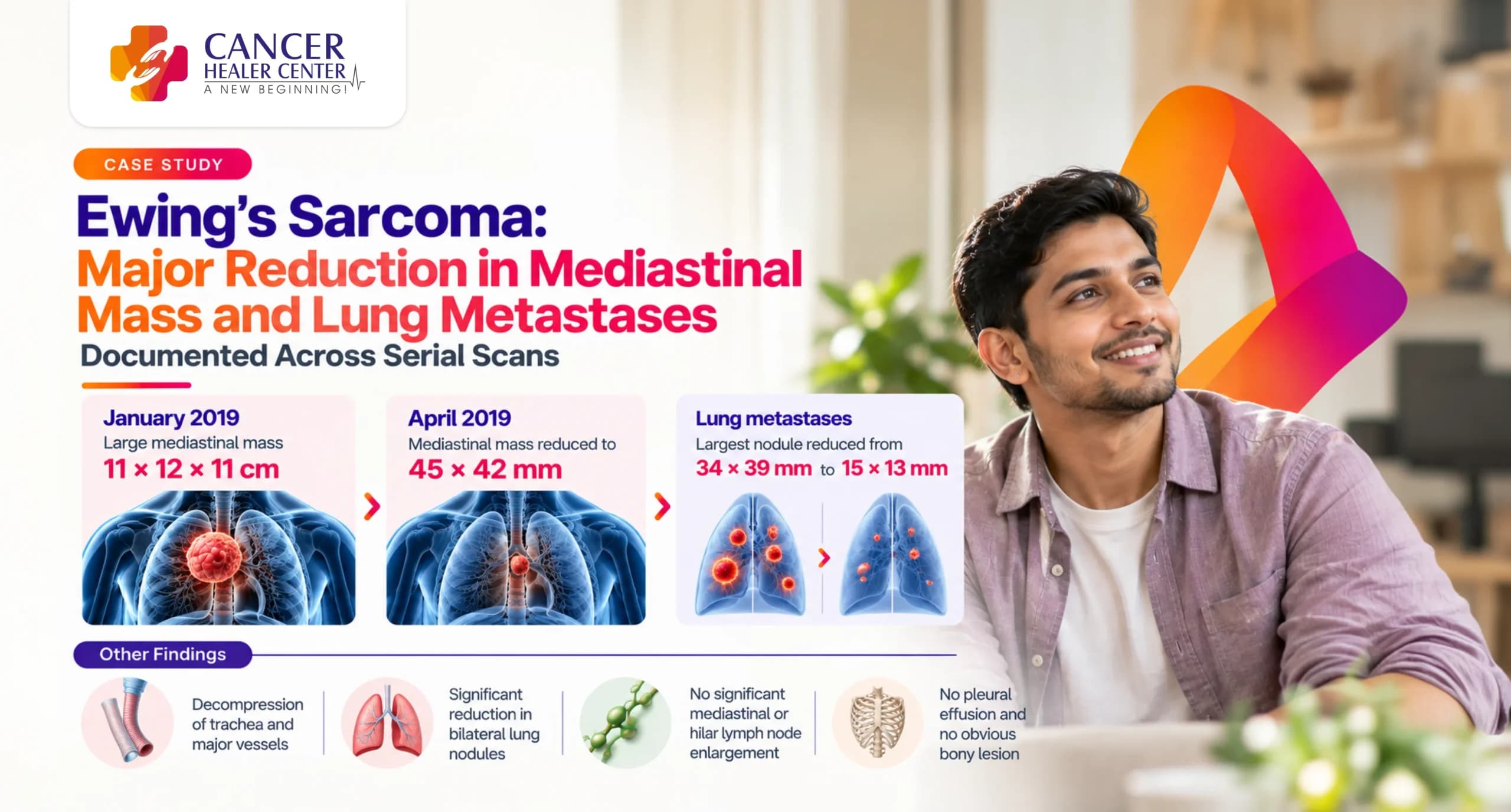
Ewing’s Sarcoma with pulmonary and mediastinal metastasis is a high-grade malignant condition requiring urgent and aggressive therapeutic intervention. This case details the clinical progress of Mr. Saddam Khan, a 24–25 year old male diagnosed with metastatic Ewing’s Sarcoma, who achieved a major objective response over a 3-month evaluation period. Serial Contrast-Enhanced Computed Tomography (CECT) scans of the thorax performed between January 2019 and April 2019 document a dramatic contraction of a massive 11 x 12 x 11 cm mediastinal tumor mass down to 45 x 42 mm, alongside substantial shrinkage and numerical reduction of bilateral pulmonary metastases.
Mr. Saddam Khan’s Case at a Glance
- Patient Name: Mr. Saddam Khan
- Age / Sex: 24–25 Years / Male
- Diagnosis: Ewing’s Sarcoma with Lung Metastases (K/c/o Ewing’s Sarcoma with lung mets)
- Primary Findings at Baseline (17/01/2019): Massive multilobulated heterogeneously enhancing soft tissue mass (~11 x 12 x 11 cm) in the right upper hemithorax and mediastinum causing mass effect on the SVC, trachea, main bronchus, and major vessels; multiple intrapulmonary and pleural nodules up to 34 x 39 mm.
- Key Scans Evaluated: CECT Scan Thorax (17/01/2019) — Bhagwan Mahaveer Cancer Hospital & Research Centre, Jaipur; CECT Thorax (27/04/2019) — Shraddha Imaging & Research Center, Sikar.
- Primary Outcome: Volumetric shrinkage of primary mediastinal tumor burden by over 80% in cross-section; decompression of major thoracic airways and vascular structures; significant reduction of bilateral lung nodules from 34 x 39 mm to 15 x 13 mm.
Mr. Saddam Khan’s Treatment Journey
Mr. Saddam Khan was diagnosed with metastatic Ewing’s Sarcoma in early 2019. Baseline high-resolution contrast-enhanced CT scanning in January 2019 revealed an advanced, life-threatening thoracic tumor burden with critical airway and vascular encasement. Under integrative treatment management with Cancer Healer Center, follow-up contrast CT imaging was conducted in April 2019 to evaluate treatment efficacy and structural disease dynamics.
What the January 2019 Baseline CT Scan Showed
A CECT Scan of the Thorax was performed on 17/01/2019 at Bhagwan Mahaveer Cancer Hospital & Research Centre, Jaipur.
Massive Mediastinal and Hilar Involvement in January 2019
- Primary Tumor Dimensions: A large, multilobulated heterogeneously enhancing soft tissue mass lesion measuring approximately 11 x 12 x 11 cm was identified in the right upper hemithorax.
- Anatomical Extension: Extended and infiltrated into the adjacent anterior, middle, and posterior mediastinum, with superior cervical extension.
- Vascular & Airway Mass Effect: Caused significant mass effect with narrowing of the right main lobe bronchus and trachea. Encasement and compression of the Superior Vena Cava (SVC), abutment of the ascending and arch of aorta, narrowing of the right main pulmonary artery and its branches, right pulmonary veins, and superior encasement of the brachiocephalic trunk and subclavian artery.
- Lymph Nodes & Collaterals: Multiple enlarged prevascular, right hilar, and subcarinal lymph nodes partially conglomerate with the thoracic mass. Multiple pleural and diaphragmatic venous collaterals were noted.
Bilateral Pulmonary and Pleural Metastases in January 2019
- Intrapulmonary & Pleural Nodules: Multiple enhancing soft tissue density pleural-based and intrapulmonary nodules across bilateral hemithoraces.
- Metastatic Nodule Sizes: The largest nodules measured 34 x 39 mm in the right middle lobe and 30 x 35 mm in the right upper lobe (perifissural).
- Pleura & Bones: No pleural or pericardial effusion was seen. No obvious bony lesion in the thoracic cage.
What the April 2019 Follow-Up CT Scan Confirmed
A repeat CECT Thorax was performed on 27/04/2019 at Shraddha Imaging & Research Center.
Significant Shrinkage of Mediastinal Tumor Mass by April 2019
- Tumor Burden Contraction: The massive 11 x 12 x 11 cm mass exhibited major regression, resolving into smaller heterogeneously enhancing lesions in the superior mediastinum, pre-aortic, right paratracheal, subcarinal, and AP window regions.
- Dimensional Reduction: The largest residual nodal mass measured only 45 x 42 mm (4.5 x 4.2 cm) in the superior mediastinum.
- Relief of Airway and Vascular Compression: Residual nodal masses produced only mild mass effect over mediastinal structures without any significant compression.
Major Reduction in Lung Metastases and Pleural Nodules
- Regression of Metastases: Only a ‘few small’ enhancing soft tissue pleural-based and intrapulmonary nodules remained in the right lung fields.
- Size Reduction: The largest residual metastatic nodule in the right lower lobe decreased from 34 x 39 mm down to approximately 15 x 13 mm. A tiny nodule remained in the left upper lobe.
Other Findings in April 2019
- Trachea & Airways: Trachea was central with a well-defined bifurcation.
- Lungs & Pleura: No interstitial septal thickening, bronchiectasis, honeycombing, or pleural effusion/thickening. Chest wall and bony thoracic cage appeared normal.
The Response Documented Over Time
| Target Region / Feature | January 2019 (Baseline CT) | April 2019 (Follow-Up CT) | Overall Clinical Response |
| Primary Mediastinal Mass | 11 x 12 x 11 cm mass with SVC & airway encasement | Largest residual mass reduced to 4.5 x 4.2 cm | Major Volumetric Reduction (>80%) |
| Airway & SVC Compression | Severe narrowing of trachea, main bronchus & SVC | Mild mass effect without significant compression | Airway & Vascular Decompression |
| Right Lung Metastases | Multiple nodules up to 34 x 39 mm & 30 x 35 mm | Few small nodules, largest 15 x 13 mm | Significant Dimensional Shrinkage |
| Left Lung Metastases | Multiple bilateral intrapulmonary nodules | Tiny single nodule in left upper lobe | Near-Complete Clearance |
| Pleural & Venous Collaterals | Multiple venous collaterals present | Resolved / Normal extra-thoracic soft tissues | Hemodynamic Improvement |
What the Two Reports Confirm
- 1. Dramatic Shrinkage of Primary Thoracic Mass: The primary tumor burden shrank from an extensive 11 x 12 x 11 cm space-occupying lesion infiltrating the entire hemithorax to localized residual nodal clusters, with the largest measuring just 45 x 42 mm.
- 2. Reversal of Critical Mass Effects: The life-threatening airway narrowing and SVC encasement observed at baseline resolved to mild displacement without significant compression.
- 3. Control of Distant Metastases: Bilateral intrapulmonary metastatic nodules underwent major regression, decreasing in maximum diameter from 39 mm to 15 mm in the right lung, while left-sided lesions cleared down to a single tiny nodule.
Understanding the CT Scan Findings
- Tumor Mass Contraction: Massive reduction in tumor dimensions indicates effective cytotoxic and cytostatic response, causing rapid cell clearance within high-grade sarcoma deposits.
- Decompression of Thoracic Structures: Relief of SVC and tracheal compression decreases the immediate risk of Superior Vena Cava Syndrome and severe airway obstruction, vastly improving respiratory mechanics and central hemodynamics.
Conclusion
This comparative evaluation reconfirms an outstanding objective response in a young male patient presenting with advanced metastatic Ewing’s Sarcoma. The rapid shrinkage of the primary thoracic mass alongside major regression of lung metastases highlights impressive structural tumor control and therapeutic efficacy.
Every Ewing’s Sarcoma Case Requires Individual Assessment
Disclaimer: Every cancer case involves unique diagnostic, histological, and anatomical factors. Treatment plans and radiological outcomes vary per individual. Clinical correlation and medical consultation remain essential for evaluating patient treatment protocols.
Medical Reports
Below are the official CT diagnostic reports documenting Mr. Saddam Khan’s clinical response:
- CECT Scan Thorax (17/01/2019) — Baseline Report (Bhagwan Mahaveer Cancer Hospital)
- CECT Thorax (27/04/2019) — Follow-Up Report (Shraddha Imaging & Research Center)
Medical Reports
Frequently Asked Questions
Mr. Saddam Khan was diagnosed with Ewing’s Sarcoma with extensive pulmonary and mediastinal metastases.
In January 2019, the primary mass in the right hemithorax measured approximately 11 x 12 x 11 cm, encasing major blood vessels (SVC, aorta, pulmonary arteries) and compressing the main bronchus and trachea.
By April 2019, the massive 11 x 12 x 11 cm tumor shrank significantly into residual nodal deposits, with the largest measuring only 45 x 42 mm (4.5 x 4.2 cm), relieving critical vascular and airway compression.
Bilateral pulmonary and pleural metastases showed marked reduction; the largest nodule shrank from 34 x 39 mm down to 15 x 13 mm, with left lung lesions resolving to a single tiny nodule.