

A Strong Response Documented Across Follow-Up Scans
Ductal carcinoma of the breast requires accurate histopathological profiling and precise ultrasonographic monitoring to track structural lesion dynamics. This case details the evaluation and monitoring of Mrs. Saraswati Mondal, a 33–34 year old female presenting with a left breast mass. Biopsy confirmed Ductal Carcinoma In Situ (DCIS) with positive hormone receptor status (ER & PR Positive). Serial ultrasonography (USG) performed between March 2020 and August 2020 documented structural stability, well-demarcated lesion boundaries, and absence of aggressive local infiltration or suspicious lymph node involvement.
Mrs. Saraswati Mondal’s Case at a Glance
- Patient Name: Mrs. Saraswati Mondal
- Age / Sex: 33–34 Years / Female
- Diagnosis: Ductal Carcinoma in Situ (DCIS) of Left Breast / Hormone Receptor-Positive Breast Carcinoma
- Biopsy & IHC Status (March 2019): Tru-cut biopsy showing moderately large pleomorphic atypical ductal cells with marked stroma sclerosis and desmoplasia. ER: Positive (Quick Score 4/8), PR: Positive (Quick Score 3/8), HER2-Neu: Negative.
- Key Investigations Evaluated: Histopathology Report (19/03/2019) — Insight Diagnostic Centre; IHC Report (29/03/2018 / 2019) — Insight Diagnostic Centre; USG Both Breasts (10/03/2020) — Apple Medical Services; USG Both Breasts (28/08/2020) — Apple Medical Services.
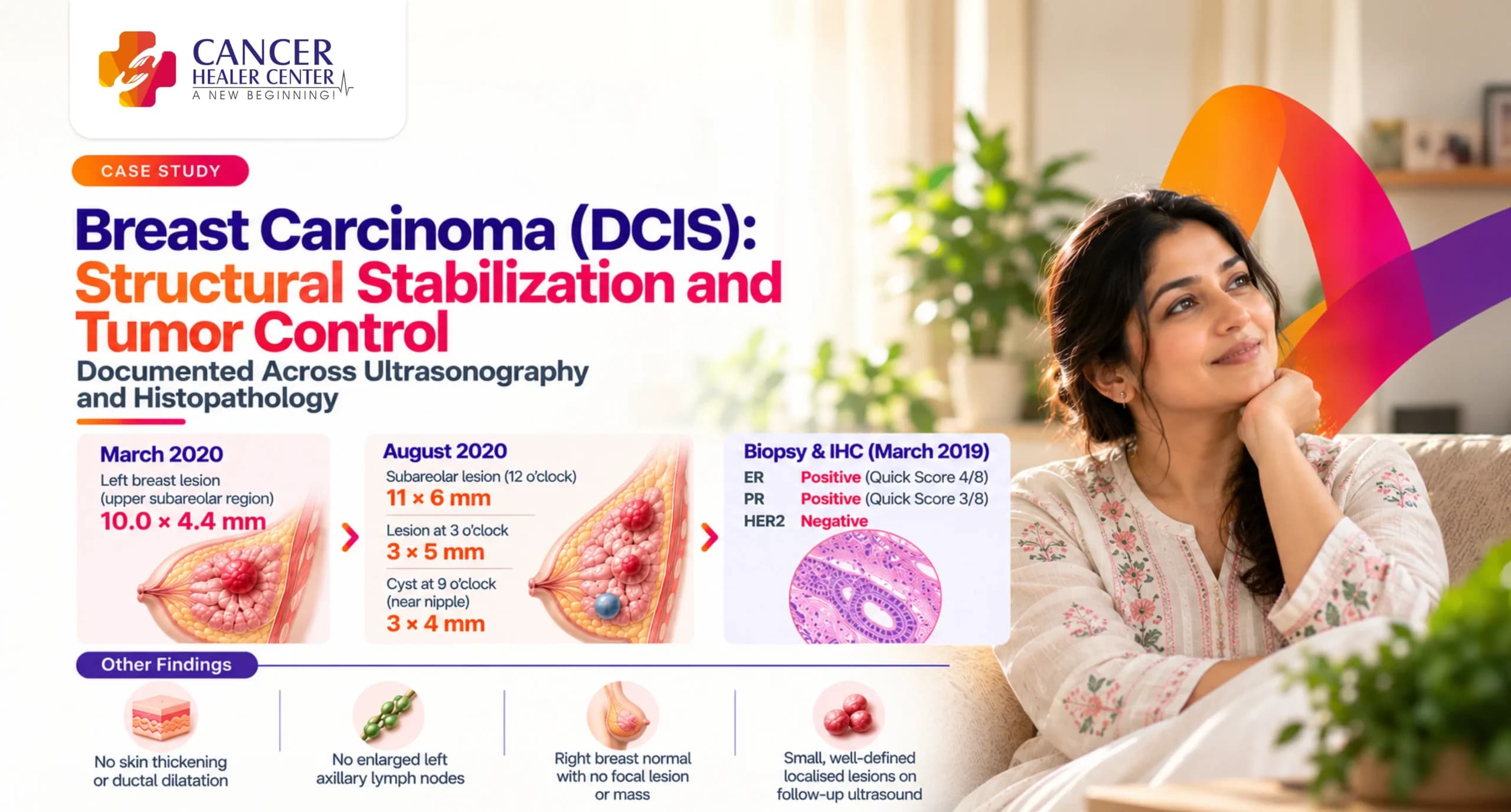
- Primary Outcome: Structural lesion stability on high-resolution sonography (10.0 x 4.4 mm in March 2020 vs localized subareolar / 3 o’clock nodules in August 2020); absence of skin thickening, ductal dilatation, or malignant axillary lymphadenopathy.
Mrs. Saraswati Mondal’s Treatment Journey
Mrs. Saraswati Mondal presented in March 2019 with a palpable mass in her left breast. Histopathological examination confirmed ductal carcinoma in situ with hormone receptor positivity (ER/PR positive, HER2 negative). Under integrative care managed through Cancer Healer Center, longitudinal monitoring via serial breast ultrasonography was conducted across 2020 to track lesion dimensions, parenchymal integrity, and axillary nodal involvement.
What the March 2019 Histopathology and IHC Showed
Diagnostic biopsy and biomarker analysis were conducted in March 2019 at Insight Diagnostic Centre, Kolkata.
Histopathology Findings in March 2019
- Specimen: True-cut biopsy from left breast mass (four linear tissue pieces).
- Microscopic Features: Breast parenchyma with fibro-collagenous and fibro-adipose tissue. Lobular architecture partly retained with distortion and distension by moderately large pleomorphic atypical ductal cells. Adjacent stroma showed marked sclerosis and desmoplasia.
- Impression: Ductal carcinoma in situ of breast in the biopsy specimen.
Immunohistochemistry (IHC) Profile in March 2019
- Estrogen Receptor (ER): Positive (Clone SP-1, Quick Score 4/8).
- Progesterone Receptor (PR): Positive (Clone EP2, Quick Score 3/8).
- HER2-Neu Status: Negative.
- Biological Subtype: Luminal A-like (Hormone Receptor Positive, HER2 Negative).
What the March 2020 Follow-Up USG Showed
A high-resolution bilateral breast ultrasound was conducted on 10/03/2020 at Apple Medical Services, Kolkata.
Baseline Ultrasonography Results (Left & Right Breast)
- Right Breast: Normal parenchymal echopattern. No focal lesion or mass seen. Skin thickness and ducts normal. Few non-specific axillary lymph nodes noted.
- Left Breast: Normal parenchymal echopattern. A small hypoechoic space-occupying lesion (SOL) measuring 10.0 x 4.4 mm identified in the upper subareolar region. No skin thickening or ductal dilatation. Few non-specific axillary lymph nodes noted.
Impression of the March 2020 USG Report
Small hypoechoic SOL (10.0 x 4.4 mm) in upper subareolar region of left breast. Bilateral axillary lymph nodes were non-specific and advised for clinical correlation.
What the August 2020 Follow-Up USG Confirmed
A follow-up bilateral breast ultrasound was conducted on 28/08/2020 at Apple Medical Services, Kolkata.
Follow-Up Ultrasonography Findings (August 2020)
- Right Breast: Normal parenchymal echopattern. No focal lesion or SOL. Axillary lymph nodes measured 6 x 6 mm (non-specific/reactive).
- Left Breast: Normal parenchymal background. Small hypoechoic SOL (3 x 5 mm) at 3 o’clock position; small clear cyst (3 x 4 mm) at 9 o’clock position near nipple; small subareolar SOL (11 x 6 mm) at 12 o’clock position. No skin thickening or ductal dilatation. Left axillary lymph nodes not enlarged.
Structural Tumor Dynamics & Node Assessment
- Impression: BI-RADS 4b — Small hypoechoic lesions in left breast with non-specific / infective bilateral axillary lymphadenopathy.
- Clinical Stability: The primary subareolar lesion remained small (11 x 6 mm) with well-defined non-invasive sonographic characteristics and no suspicious lymph node structural transformation.
The Response Documented Over Time
| Diagnostic Parameter | March 2019 (Biopsy / IHC) | March 2020 USG | August 2020 USG | Clinical Outcome |
| Left Breast Mass Size | Palpable mass (Biopsy positive DCIS) | 10.0 x 4.4 mm subareolar SOL | 11 x 6 mm subareolar SOL | Controlled & Stable Dimensions |
| Parenchymal & Skin Features | Sclerosis & desmoplasia on biopsy | Normal skin thickness & non-dilated ducts | No skin thickening or ductal dilatation | No Local Tissue Infiltration |
| Axillary Lymph Nodes | Not evaluated on biopsy | Few non-specific axillary nodes | Non-specific / benign (6 x 6 mm) | No Malignant Lymphadenopathy |
| Hormone Status / Subtype | ER+ / PR+ / HER2- | N/A | N/A | Favorable Luminal A Profile |
What the Serial Reports Confirm
- 1. Hormone-Sensitive Biology: IHC profiling confirms an ER/PR positive, HER2-negative tumor signature suitable for endocrine and targeted metabolic regulation.
- 2. Sonographic Stability: Serial ultrasound monitoring confirms stable, small localized nodular dimensions (10.0 x 4.4 mm vs 11 x 6 mm) without aggressive local expansion, skin tethering, or suspicious axillary node conversion.
Understanding the Histopathology and USG Findings
- DCIS & Desmoplasia: Histology identified distorted ductal architecture with pleomorphic atypical cells and stromal desmoplasia. High-resolution ultrasound tracks whether these micro-architectural changes extend into surrounding tissue.
- Hormone Receptor Positivity: Positive ER and PR scores (Quick Score 4/8 and 3/8) indicate hormone responsiveness and favorable clinical prognosis compared to receptor-negative subtypes.
Conclusion
This serial diagnostic review confirms clinical stability and localized structural control in a young patient with hormone receptor-positive left breast ductal carcinoma in situ. Serial ultrasonography demonstrates controlled lesion dimensions without invasive anatomical spread or malignant axillary nodal progression.
Every Breast Carcinoma Case Requires Individual Assessment
Disclaimer: Every cancer case involves unique diagnostic, histological, and anatomical factors. Treatment plans and radiological outcomes vary per individual. Clinical correlation and medical consultation remain essential for evaluating patient treatment protocols.
Medical Reports
Below are the official diagnostic reports documenting Mrs. Saraswati Mondal’s clinical evaluation:
- Histopathology Report (19/03/2019) — Left Breast Biopsy (Insight Diagnostic Centre)
- Immunohistochemistry Report (29/03/2018 / 2019) — ER / PR / HER2 (Insight Diagnostic Centre)
- USG Both Breasts Report (10/03/2020) — Baseline USG (Apple Medical Services)
- USG Both Breasts Report (28/08/2020) — Follow-Up USG (Apple Medical Services)
Medical Reports
Frequently Asked Questions
The tru-cut biopsy performed in March 2019 confirmed Ductal Carcinoma In Situ (DCIS) of the left breast.
The IHC test confirmed ER Positive (Quick Score 4/8), PR Positive (Quick Score 3/8), and HER2-Neu Negative, indicating a hormone receptor-positive breast carcinoma.
The ultrasounds performed in March 2020 and August 2020 confirmed a small, well-defined hypoechoic subareolar lesion measuring 10.0 x 4.4 mm (March) and 11 x 6 mm (August), with normal skin thickness and no suspicious axillary lymph nodes.
No. Both ultrasounds showed normal parenchymal backgrounds with only non-specific, reactive axillary lymph nodes.