
A Strong Response Documented Across Follow-Up Scans
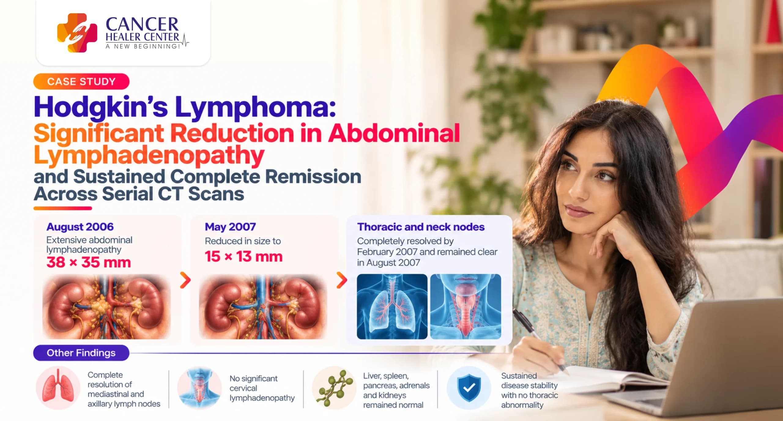
Hodgkin’s Lymphoma recurrence presents complex clinical challenges requiring meticulous longitudinal monitoring. This case details the evaluation of Mrs. Rekha, a young female diagnosed with a recurrence of Hodgkin’s Lymphoma, who demonstrated a remarkable response over a 12-month evaluation period. Serial Computed Tomography (CT) scans performed between August 2006 and August 2007 demonstrate complete resolution of early mediastinal and axillary thoracic lesions alongside a significant reduction in extensive abdominal lymphadenopathy.
Mrs. Rekha’s Case at a Glance
- Patient Name: Mrs. Rekha Rani (Mrs. Rekha)
- Age / Sex: 26–28 Years / Female
- Diagnosis: Recurrent Hodgkin’s Lymphoma (Primary diagnosis in 1999)
- Primary Findings at Recurrence (August 2006): Enlarged right pretracheal, subcarinal, left axillary, pre/para-aortic, aortocaval, retrocaval, and bilateral iliac lymph nodes
- Key Scans Evaluated: CT Neck, Chest, and Whole Abdomen (17/08/2006); CECT Chest (12/02/2007); CT Scan of Abdomen (13/02/2007); CECT Neck (11/05/2007); CECT Chest (11/05/2007); CT Scan of Abdomen (11/05/2007); CECT Chest (09/08/2007)
- Primary Outcome: Total clearance of mediastinal, axillary, and neck involvement; major dimension reduction in retroperitoneal and para-aortic lymph nodes (from 38 x 35 mm down to 15 x 13 mm); sustained disease stability.
Mrs. Rekha’s Treatment Journey
Mrs. Rekha originally underwent treatment for Hodgkin’s Lymphoma in 1999. In August 2006, at age 27, she reported for evaluation of disease recurrence. Baseline imaging confirmed recurrent lymphadenopathy across thoracic and extensive abdominal nodal stations. Following integrative therapy management under Cancer Healer Center, follow-up contrast-enhanced CT scans were systematically conducted over the course of 2007 to monitor structural disease dynamics and anatomical response.
What the August 2006 Baseline CT Scan Showed
A comprehensive CT examination of the neck, chest, and whole abdomen was performed on 17/08/2006 at Rajiv Gandhi Cancer Institute and Research Centre.
Chest and Neck Assessment in August 2006
- Neck: Nasopharynx, oropharynx, hypopharynx, larynx, thyroid, and salivary glands were normal. No significant cervical lymphadenopathy was observed.
- Chest Nodes: Enlarged right pretracheal lymph node, subcarinal lymph node, and left axillary lymph nodes were present. Soft-tissue thickening was identified in the prevascular region.
- Lungs & Pleura: Both lungs, trachea, and main stem bronchi were normal with no pleural or pericardial effusion.
Abdominal Findings in August 2006
- Retroperitoneal Nodes: Extensive enlargement of confluent and discrete lymph nodes starting from the celiac axis level. Pre-aortic, para-aortic, aortocaval, retrocaval, and common iliac nodes displaced the celiac artery and renal vessels, extending down to the iliac vessel bifurcation. Bilateral external iliac nodes were also enlarged.
- Abdominal Organs: Liver, gallbladder, spleen, pancreas, adrenals, and kidneys were normal.
What the February 2007 Follow-Up CT Scans Showed
Six months into evaluation, dedicated CT imaging of the chest and abdomen was performed on 12/02/2007 and 13/02/2007 at Shri Anandpur Trust Charitable Diagnostic Centre.
Complete Resolution of Chest Abnormality in February 2007
CECT Chest (12/02/2007) showed complete clearance of previously noted right pretracheal, subcarinal, and left axillary lymph nodes as well as prevascular soft tissue thickening. Impression: ‘NO SIGNIFICANT ABNORMALITY SEEN.’
Abdominal Scan Results in February 2007
CT Abdomen (13/02/2007) showed significantly enlarged discrete and confluent lymph nodes remaining in the para-aortic region and along common iliac vessels, measuring 38 x 35 mm in maximum dimensions. No renal mass, hydronephrosis, or focal liver/spleen lesions were present.
What the May 2007 Follow-Up CT Scans Confirmed
Follow-up examinations conducted on 11/05/2007 provided objective evidence of substantial regressive changes.
Normal Neck Scan in May 2007
CECT Neck (11/05/2007): Normal structures throughout. No evidence of significant lymphadenopathy. Impression: ‘NO SIGNIFICANT ABNORMALITY SEEN.’
Sustained Normal Chest Findings in May 2007
CECT Chest (11/05/2007): Reconfirmed clear lung parenchyma, well-maintained mediastinal fat planes, and normal hila without lymphadenopathy. Impression: ‘NO DEFINITE ABNORMALITY SEEN.’
Significant Structural Reduction in Abdominal Nodes by May 2007
CT Abdomen (11/05/2007): Persistent but substantially reduced discrete and confluent lymph nodes in the para-aortic, aortocaval, common iliac, and left external iliac regions. The maximum nodal size decreased significantly from 38 x 35 mm (February 2007) down to 15 x 13 mm (May 2007) in the left para-aortic region. No evidence of calcification or necrosis was observed.
What the August 2007 Chest Scan Confirmed
A repeat CECT Chest on 09/08/2007 confirmed long-term stability and complete absence of thoracic disease re-emergence. Lungs, pleura, and mediastinum remained clear with intact fat planes and normal hila. Impression: ‘NO DEFINITE ABNORMALITY SEEN. NO INTERVAL CHANGE PREVIOUS SCAN DATE 11-05-2007.’
The Response Documented Over Time
| Target Region / Feature | August 2006 (Baseline) | Feb 2007 Scan | May 2007 Scan | Aug 2007 Scan |
| Pretracheal & Subcarinal Nodes | Enlarged | Completely Resolved | Completely Resolved | Completely Resolved |
| Left Axillary Nodes | Enlarged | Completely Resolved | Completely Resolved | Completely Resolved |
| Prevascular Soft Tissue | Soft tissue thickening present | Resolved | Resolved | Resolved |
| Para-aortic / Abdominal Nodes | Confluent, extensive enlargement | Max size 38 x 35 mm | Reduced to 15 x 13 mm | Not scanned (Chest stable) |
| Neck Lymph Nodes | No significant lymphadenopathy | Normal | Completely Normal | Normal |
What the Serial Reports Confirm
The comparative radiological findings confirm a structured, sequential response to therapy:
- 1. Rapid Clearance of Thoracic Disease: Early follow-up in February 2007 confirmed total resolution of pretracheal, subcarinal, prevascular, and left axillary nodal disease, which remained clear through August 2007.
- 2. Marked Contraction of Abdominal Disease: Retroperitoneal lymphadenopathy demonstrated a dramatic volumetric shrinkage, reducing in maximum axial cross-section from 38 x 35 mm to 15 x 13 mm within three months between February and May 2007.
Understanding the CT Scan Findings
- Lymphadenopathy Resolution: The regression of enlarged lymph nodes indicates structural control and cellular clearance of malignant disease within nodal basins.
- Interval Stability: Maintaining clear scan findings across consecutive evaluations (May 2007 to August 2007) validates sustained remission without acute interval progression or relapsing thoracic disease.
Conclusion
This serial CT evaluation confirms an outstanding therapeutic response in a young patient presenting with recurrent Hodgkin’s Lymphoma. The rapid resolution of thoracic disease combined with a greater than 60% reduction in maximal retroperitoneal nodal dimensions underscores significant treatment efficacy and sustained clinical stability.
Every Hodgkin’s Lymphoma Case Requires Individual Assessment
Disclaimer: Every cancer case involves unique diagnostic, histological, and anatomical factors. Treatment plans and radiological outcomes vary per individual. Clinical correlation and medical consultation remain essential for evaluating patient treatment protocols.
Medical Reports
Below is the record of official CT scan diagnostic reports documenting Mrs. Rekha’s clinical progression:
- CT Scan Report (17/08/2006) — Baseline Neck, Chest & Abdomen (Rajiv Gandhi Cancer Institute)
- CT Scan Report (13/02/2007) — Follow-up Abdomen (Shri Anandpur Trust)
- CT Scan Report (11/05/2007) — Follow-up Abdomen (Shri Anandpur Trust)
- CECT Chest Report (12/02/2007) — Follow-up Chest (Shri Anandpur Trust)
- CECT Neck Report (11/05/2007) — Follow-up Neck (Shri Anandpur Trust)
- CECT Chest Report (11/05/2007) — Follow-up Chest (Shri Anandpur Trust)
- CECT Chest Report (09/08/2007) — Follow-up Chest (Shri Anandpur Trust)
Medical Reports
Frequently Asked Questions
In August 2006, the CT scan showed involvement in the chest (right pretracheal, subcarinal, left axillary nodes, and prevascular soft tissue) as well as extensive abdominal involvement (pre-aortic, para-aortic, aortocaval, retrocaval, and common/external iliac lymph nodes).
By February 2007 (6 months after baseline imaging), the chest CT confirmed complete resolution of all mediastinal and axillary lymphadenopathy.
The retroperitoneal lymph nodes decreased from 38 x 35 mm in February 2007 to 15 x 13 mm in May 2007, reflecting significant tumor burden reduction.
No. The August 2007 CECT Chest scan confirmed no definite abnormality and no interval changes compared to May 2007, demonstrating stable complete remission in the thoracic cavity.