

A PET-CT scan is one of the most useful imaging tests in cancer care. It is also one of the most confusing for patients. It involves an injection, a long wait and a machine you may never have seen before. Knowing what to expect makes the day much easier.
This guide explains what a PET-CT scan for cancer is, when it is used, how to prepare, what happens during the procedure, how to understand the report and what affects the cost in India. It cannot replace advice from your own doctor.
Have a question about a scan your doctor has advised?
What Is a PET-CT Scan?
PET stands for positron emission tomography. A PET scan uses a tiny amount of a radioactive tracer that is injected into a vein. The most common tracer is FDG, a form of sugar. Cancer cells often use more sugar than normal cells, so they collect more tracer, and a special camera picks this up as bright areas.
A PET scan alone does not show exact anatomy. That is why it is almost always done on a combined machine. The CT part takes detailed X-ray pictures of the body, and the PET part adds the activity. Together, the PET CT scan shows both what and where (Fletcher et al., 2008).
What Is a PET Scan Used For in Cancer?
- Staging. It shows whether a cancer has spread to lymph nodes or other organs. This helps decide the treatment.
- Checking response. It shows whether a tumour is shrinking or becoming less active during or after chemotherapy or radiation. In lymphoma, response is scored on PET using a standard system (Cheson et al., 2014).
- Finding a recurrence. It helps look for a return of cancer when symptoms appear or a tumour marker is rising.
- Finding the source. It can help look for the original site when cancer is found in a lymph node or distant organ.
- Planning treatment. Radiation teams use it to outline the target, and surgeons use it to plan an operation.
A PET scan is also not meant for everyone. It is not used to screen healthy people, and it is not always the right test for small, slow-growing tumours.
Different tracers for different cancers
| Tracer | Used mainly for |
|---|---|
| FDG | Most cancers, including lung, lymphoma, head and neck, oesophagus, colon and breast |
| PSMA | Prostate cancer, especially when it returns after treatment. A trial showed it was more accurate than standard imaging for staging (Hofman et al., 2020) |
| DOTATATE | Neuroendocrine tumours. |
PET-CT Scan Preparation
Preparation instructions differ slightly between centres, so follow those you are given. In general (Boellaard et al., 2015):
- Fast for four to six hours. You can drink plain water, and drinking water helps. Do not have tea, coffee with sugar, juice or a sugary drink.
- Avoid heavy exercise for about 24 hours before, because muscle activity can show up on the scan.
- Tell the centre if you have diabetes. High blood sugar can reduce the quality of the scan. Most centres want a level below about 200 mg/dL, and they will tell you how to manage your tablets or insulin.
- Bring your reports and earlier scans. The doctor reading the scan needs them for comparison, along with your list of medicines.
- Wear loose, metal-free clothing, and leave jewellery and belts at home.
- Tell the staff if you are pregnant or breastfeeding, or if you are afraid of closed spaces.
- Ask about timing. If you recently had chemotherapy, radiation or a biopsy, your doctor may time the scan to avoid misleading results.
What Happens During a PET-CT Scan
The whole visit usually takes two to three hours, though the scan itself is short.
- Check-in. Staff check your details, weight, reports and blood sugar with a small finger prick.
- Injection. The tracer is injected into a vein in your arm. You will not feel anything unusual.
- Uptake period. You rest quietly in a room for about 45 to 60 minutes while the tracer spreads. Try not to talk much, read or use your phone heavily, because muscle activity can affect the images. You may be asked to empty your bladder before the scan.
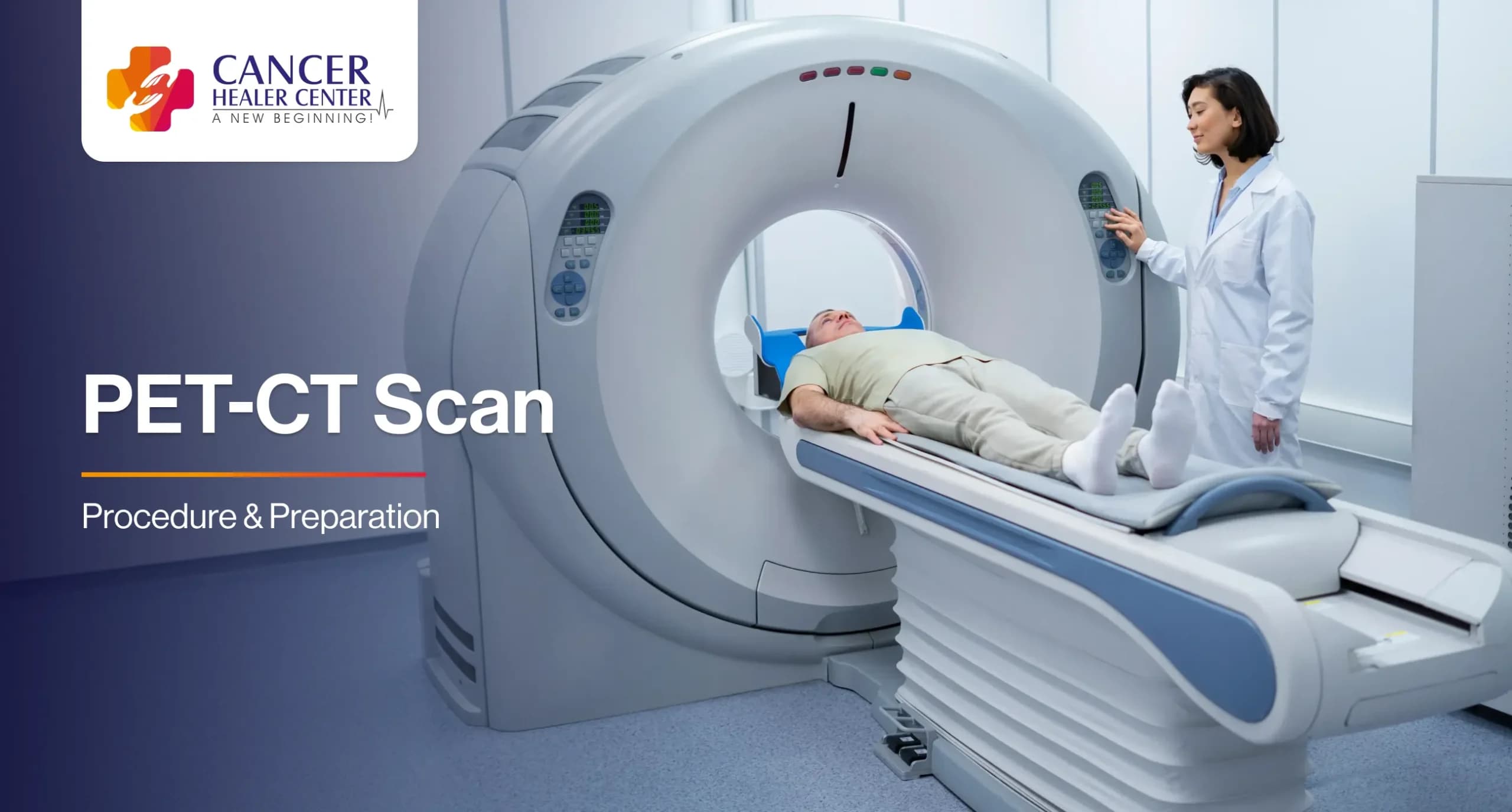
- Scan. You lie on a narrow table that slides slowly through a ring-shaped machine. It takes about 20 to 30 minutes. It is painless and not as enclosed as an MRI. You need to stay still.
- After the scan. You can usually eat, drink and go home. Drinking more fluid helps flush out the tracer.
Sometimes the CT part uses contrast dye, given by injection or as a drink. You will be told if this is planned, and asked about allergies and kidney problems.
Is a PET-CT scan safe?
The amount of radiation is small, and the tracer fades within a day. The benefit of getting the right information about cancer usually outweighs the small risk. For a few hours afterwards, staff may advise you to keep a distance from infants and pregnant women. PET-CT is generally avoided in pregnancy unless it is essential, and breastfeeding mothers get specific advice from the centre.
How to Read a PET-CT Report
Your report may use words such as “avid”, “hypermetabolic” or SUV, a number that measures how much tracer an area took up. A higher value means more activity. It does not on its own mean cancer.
Bright spots can also appear with infection, inflammation, tuberculosis, a healing wound, a recent biopsy, recent radiation, or normal activity in the brain, heart, muscles and bladder. In the same way, very small tumours or slow-growing tumours may not light up at all. So the PET result is always read together with the CT, your symptoms and the biopsy.
PET-CT Scan Cost in India
People often search for the PET scan cost. There is no single price, because the charge depends on several things:
| What affects the price | Why it matters |
|---|---|
| City and type of centre | Metro hospitals, standalone diagnostic centres and government hospitals charge differently |
| Tracer used | FDG is the most common. PSMA and DOTATATE tracers usually cost more |
| Area scanned | A whole-body scan costs more than a limited scan |
| Contrast | CT contrast may be charged extra |
| Reporting and extras | Some centres include the report, CD and consultation, others charge separately |
Before you book, ask the centre for the current charge, what it includes and whether there are extra charges for contrast or a second review. Some insurance policies and government schemes cover PET-CT when a doctor advises it for cancer, subject to their terms, so check with your insurer or scheme about approval before the scan.
Cost should not be the only thing you compare. A centre with a good machine, an experienced nuclear medicine doctor and a quick report is worth more than a small difference in price. Also, ask whether your doctor really needs the scan now. An unnecessary scan costs money and can cause worry if it picks up harmless findings.
How PET-CT Fits With Other Cancer Tests
A PET-CT scan is one piece of the diagnostic puzzle. A biopsy confirms what the cells are, blood tests such as tumour markers give extra clues, and the PET-CT shows how far the disease has spread.
When Should You See a Doctor?
Talk to your doctor if you have a lump that keeps growing, unexplained weight loss, a long-lasting fever, night sweats, persistent pain, bleeding, a cough that will not clear or any symptom that has lasted more than a few weeks. Do not book a PET scan on your own. It should be advised by your treating doctor for a clear reason.
After the scan, if you have a rash, swelling at the injection site or breathing difficulty after contrast dye, tell the centre or go to an emergency department.
Support at Cancer Healer Center
Cancer Healer Center offers consultations for people with cancer and their families, including remote consultations for those who cannot travel. Please bring all your scans and reports with you. The centre also offers Cancer Healer Therapy (CAM), a herbal-based supportive approach meant to be used alongside the treatment prescribed by your oncologist and not instead of it.
For appointments or general enquiries, reach out to the centre.
References
Boellaard, R., Delgado-Bolton, R., Oyen, W.J.G., et al. (2015) ‘FDG PET/CT: EANM procedure guidelines for tumour imaging: version 2.0’, European Journal of Nuclear Medicine and Molecular Imaging, 42(2), pp. 328-354. doi:10.1007/s00259-014-2961-x.
Cheson, B.D., Fisher, R.I., Barrington, S.F., et al. (2014) ‘Recommendations for initial evaluation, staging, and response assessment of Hodgkin and non-Hodgkin lymphoma: the Lugano classification’, Journal of Clinical Oncology, 32(27), pp. 3059-3068. doi:10.1200/JCO.2013.54.8800.
Fletcher, J.W., Djulbegovic, B., Soares, H.P., et al. (2008) ‘Recommendations on the use of 18F-FDG PET in oncology’, Journal of Nuclear Medicine, 49(3), pp. 480-508. doi:10.2967/jnumed.107.047787.
Hofman, M.S., Lawrentschuk, N., Francis, R.J., et al. (2020) ‘Prostate-specific membrane antigen PET-CT in patients with high-risk prostate cancer before curative-intent surgery or radiotherapy (proPSMA): a prospective, randomised, multicentre study’, The Lancet, 395(10231), pp. 1208-1216. doi:10.1016/S0140-6736(20)30314-7.
Frequently Asked Questions
A PET scan is an imaging test that uses a small amount of radioactive tracer to show how active the body’s cells are. Cancer cells often take up more tracer and appear as bright areas.
A PET-CT scan combines the PET scan and a CT scan in one machine. The CT part shows the body’s structure, and the PET part shows activity, so doctors can see exactly where an active area is.
Fast for four to six hours, drink plain water, avoid heavy exercise for a day, tell the centre about diabetes, pregnancy and allergies, and bring your reports. Follow the centre’s own instructions.
The visit takes about two to three hours. The injection and a rest period of about an hour come first, then the scan itself takes 20 to 30 minutes.
No. You feel only the needle for the injection. You then lie still on the table while the scan is taken.
No. It can miss small or slow-growing tumours and some cancers do not take up the tracer well. It can also light up infection or inflammation. A biopsy is still needed to confirm cancer.
The cost depends on the city, the centre, the tracer and the area scanned. Ask the centre for the current charge and what it includes, and check with your insurer or scheme.