

When a doctor suspects cancer, two words come up again and again: biopsy and PET scan. They sound technical, and many people are frightened by them. Knowing what each test is for, what happens on the day and what the results can and cannot tell you makes the process much less stressful.
This guide explains what a biopsy is, the common types, how a PET CT scan works, how to prepare and how doctors use both tests to diagnose and stage cancer. It cannot replace advice from your own doctor.
Have a question about a test your doctor has advised?
What Is a Biopsy?
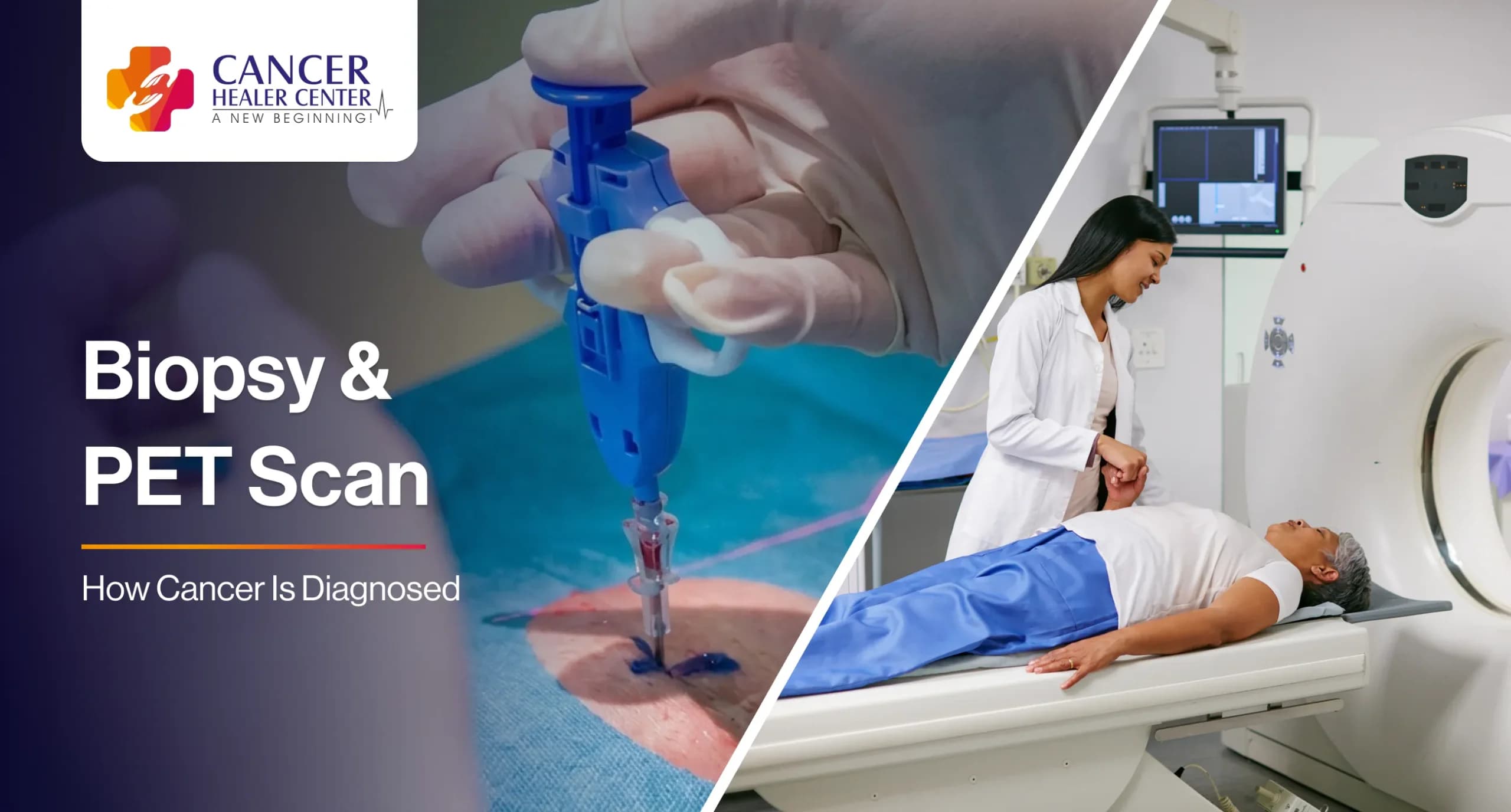
Biopsy means a procedure in which a small sample of tissue or cells is taken from the body and studied by a pathologist under a microscope. If you are asking what is a biopsy test, it is the standard way to confirm a cancer diagnosis, because scans and blood tests can only suggest that something is wrong. They cannot show what kind of cells are present.
A biopsy report can tell you:
- Whether the growth is benign (not cancer) or malignant (cancer)
- The type of cancer and how fast the cells are likely to grow, called the grade
- Whether special features are present, such as hormone receptors in breast cancer, which guide treatment
- In surgical samples, whether the edges of the removed tissue are clear
Types of biopsy test
| Type | How it is done | Common uses |
|---|---|---|
| Fine needle aspiration (FNAC) | A thin needle removes a few cells | Thyroid, neck nodes, breast lumps |
| Core needle biopsy | A slightly larger needle removes a thin core of tissue, often under ultrasound or CT guidance | Breast, liver, lung, prostate, soft tissue |
| Endoscopic biopsy | A camera tube takes samples from inside the gut, airway or bladder | Stomach, food pipe, colon, lung, bladder |
| Incisional biopsy | A small cut removes part of a lump | Mouth, skin, soft tissue and bone tumours |
| Excisional biopsy | The whole lump is removed | Small skin growths, lymph nodes |
| Bone marrow biopsy | A needle takes marrow from the hip bone | Blood cancers such as leukaemia and lymphoma |
Some people worry that a biopsy can spread cancer. The risk is very low, and doctors plan the route of the needle or cut to reduce it further. Not having a biopsy and delaying diagnosis carries a bigger risk.
How to prepare and what to expect
Tell the doctor about blood thinners, aspirin, allergies, diabetes and pregnancy. You may be asked to fast or to stop certain medicines for a short time. Most needle biopsies are done under local anaesthesia and take 15 to 30 minutes. Endoscopic and surgical biopsies may need sedation or general anaesthesia. Afterward you may feel soreness or have a little bruising. Serious problems such as heavy bleeding or infection are uncommon, but call your doctor if you develop fever, increasing pain, swelling or bleeding.
Results usually take several days. Extra tests on the tissue, such as markers or gene studies, may take a week or more.
What Is a PET Scan?
A PET scan, short for positron emission tomography, is an imaging test that shows how active the cells in the body are. Most cancer PET scans use a form of sugar tagged with a tiny amount of radioactive tracer, called FDG. Cancer cells often use more sugar than normal cells, so they take up more of the tracer and show up as bright spots on the images (Fletcher et al., 2008).
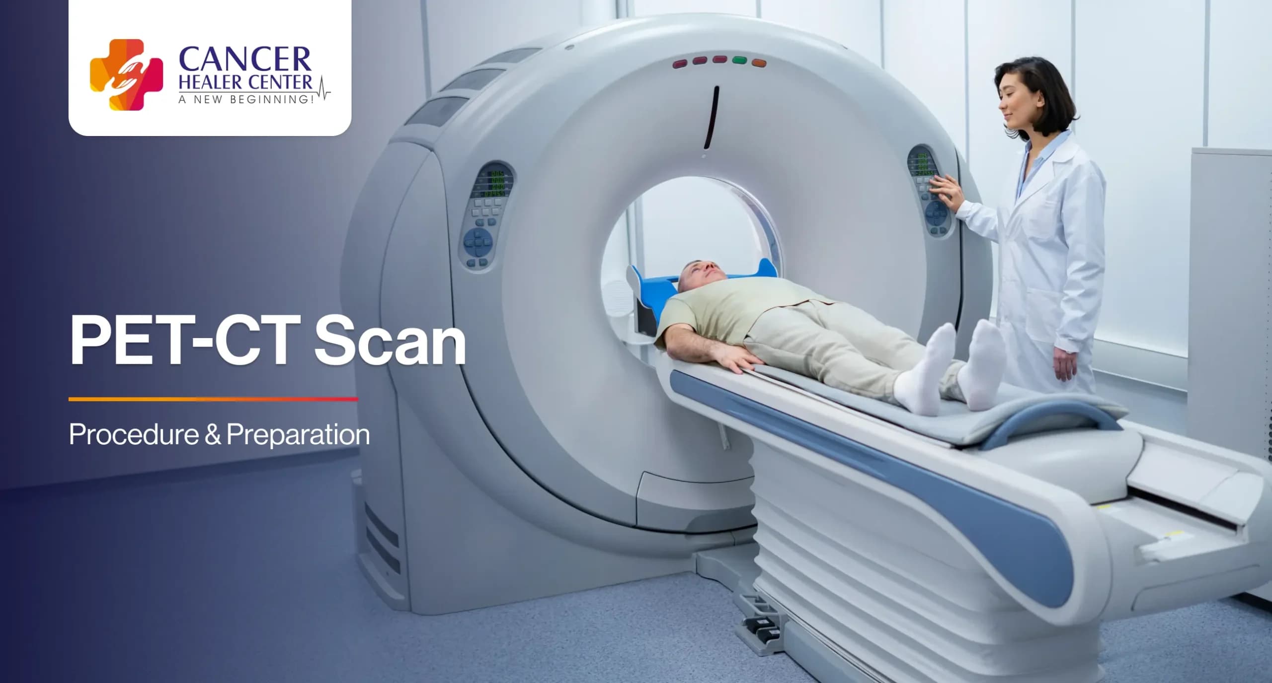
Today most scans are done on a combined machine, a PET CT scan or PET-CT. The CT part gives a detailed picture of the body’s structure, and the PET part adds information on activity. Together they show exactly where the active area sits.
PET Scan for Cancer
A PET scan for cancer helps doctors in several ways (Fletcher et al., 2008):
| Purpose | Description |
|---|---|
| Staging | It checks whether a known cancer has spread to lymph nodes or distant organs. |
| Checking response | It shows whether a tumour is responding to chemotherapy or radiation. |
| Finding a return | It helps look for recurrence when a tumour marker is rising or symptoms return. |
| Finding the source | It can help look for the original site when cancer is found in a node or distant organ but the starting point is unknown. |
| Planning treatment | It helps radiation teams and surgeons outline the area to treat. |
PET is not the best test for every cancer. Some slow-growing tumours take up little FDG, and other tracers are used instead. For example, a PSMA tracer is used for prostate cancer and a DOTATATE tracer for neuroendocrine tumours.
How to Prepare for a PET CT Scan and What Happens
Preparation instructions vary between centres, so follow the ones you are given. In general (Boellaard et al., 2015):
- Fasting. You usually stop eating for four to six hours before the scan. You can drink plain water.
- Avoid heavy exercise for about a day before.
- Diabetes. Tell the centre if you have diabetes. Blood sugar levels affect the scan and may need to be managed.
- Tell the staff if you are pregnant, breastfeeding or claustrophobic.
- Injection and rest. The tracer is injected into a vein. You then rest quietly for about an hour while it spreads through the body.
- Scan. You lie on a table that slides through the machine for about 20 to 30 minutes. It is painless.
The radiation dose is small, and the tracer leaves the body within a day. Staff may advise you to keep a distance from young children and pregnant women for some hours after the scan.
PET Scan Cost in India
People often search for the PET scan cost in India. A PET CT scan costs more than a plain CT or MRI, and the price varies with the city, the centre, the tracer used and the area scanned. Ask the centre for the current charge and what it includes, such as the injection, the CT part and the report, before you book.
Biopsy vs PET Scan
| Biopsy | PET CT scan | |
|---|---|---|
| What it is | Removal of a tissue sample for study | An imaging scan with a radioactive tracer |
| What it tells you | Whether a growth is cancer, its type and grade | Where active disease may be and how far it has spread |
| Can it confirm cancer alone? | Yes, in most cases | No, it needs a biopsy to confirm |
| Main limits | Only samples the area taken | Can light up infection or inflammation, and can miss small or slow-growing tumours |
In practice, doctors often start with an ultrasound, CT or MRI that finds a suspicious area, then take a biopsy to confirm what it is, and later use a PET CT scan to stage the cancer and plan treatment. Blood tests such as tumour markers add information but cannot replace either test.
Understanding Your Results
A PET report may describe an area as “avid” or “hypermetabolic”, which means it took up a lot of tracer. This is not the same as saying it is cancer. Infections such as tuberculosis, inflammation, and healing tissue can also light up. A report that shows no uptake does not completely rule out cancer, because very small tumours can be missed. Your doctor reads the PET result together with the biopsy, the CT findings and your symptoms.
If you are waiting for results, this can be a hard time. Ask your doctor when to expect them and who will explain them to you.
When Should You See a Doctor?
See a doctor if you have a lump that is growing or does not go away, unexplained weight loss, a sore that does not heal, long-lasting changes in bowel or bladder habit, unusual bleeding, a cough that will not go away or persistent pain. After a biopsy, call your doctor if you develop fever, spreading redness, heavy bleeding or severe pain at the site.
Diagnosis Support at Cancer Healer Center
Cancer Healer Center offers consultations for people with cancer and their families, including remote consultations for those who cannot travel. Please bring your biopsy report, scans and blood tests when you consult. The centre also offers Cancer Healer Therapy (CAM), a herbal-based supportive approach meant to be used alongside the treatment prescribed by your oncologist and not instead of it.
Have questions about your care options?
References
Boellaard, R., Delgado-Bolton, R., Oyen, W.J.G., et al. (2015) ‘FDG PET/CT: EANM procedure guidelines for tumour imaging: version 2.0’, European Journal of Nuclear Medicine and Molecular Imaging, 42(2), pp. 328-354. doi:10.1007/s00259-014-2961-x.
Fletcher, J.W., Djulbegovic, B., Soares, H.P., et al. (2008) ‘Recommendations on the use of 18F-FDG PET in oncology’, Journal of Nuclear Medicine, 49(3), pp. 480-508. doi:10.2967/jnumed.107.047787.
Frequently Asked Questions
A biopsy is a procedure that removes a small sample of tissue or cells so a pathologist can study it under a microscope. It is the standard way to confirm whether a growth is cancer.
It confirms or rules out cancer, identifies the type and grade, and tests for features that guide treatment, such as hormone receptors in breast cancer.
The risk is very low. Doctors plan the route of the needle or cut to minimise it. Delaying a diagnosis is a bigger risk than having a biopsy.
A PET scan is an imaging test that uses a small amount of radioactive tracer to show how active the cells in the body are. Cancer cells often take up more tracer and show up as bright areas.
A PET CT scan combines the PET scan with a CT scan in one machine. The CT part shows detailed body structure, and the PET part shows activity, so doctors can see exactly where an active area sits.
No. It can miss small or slow-growing tumours, and it can light up infection or inflammation. Results are always read along with other tests.
The price varies with the centre, city, tracer and body area scanned. Ask the centre for the current charge and what it includes before you book.