

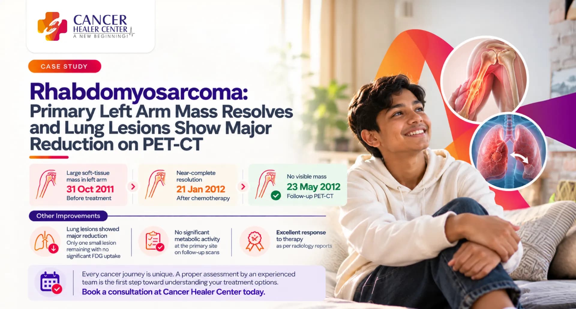
A Strong Response Documented Across Two Follow-Up Scans
Md Ashraf was 15 years old when he underwent a whole-body PET-CT to assess his response to treatment for a suspected malignant mass in the proximal left humerus. He had been receiving adjuvant chemotherapy, and an earlier scan dated 31 October 2011 was available for comparison.
The PET-CT reported on 21 January 2012 showed near-complete resolution of the large soft-tissue mass in his left upper arm. No significant FDG uptake was seen at the site. The scan also showed the virtual disappearance of the previously documented lung lesions, except for one remaining lesion in the lingular segment of the left lung. This lesion had reduced substantially and measured 29 × 25 mm, with no significant FDG uptake.
A further PET-CT reported on 23 May 2012 identified the diagnosis as rhabdomyosarcoma of the proximal left humerus. By this assessment, no soft-tissue mass was visible in the left arm, and no significant metabolic activity was present at the site of the primary lesion. The remaining left lung nodule had reduced further in size and continued to show no significant FDG uptake.
The radiologists described the overall findings as an excellent response to therapy.
Note: This case study presents Md Ashraf’s documented treatment journey and PET-CT findings. Individual outcomes may vary depending on the type and extent of cancer, previous treatment, overall health and response to therapy.
Md Ashraf’s Case at a Glance
| Patient detail | Documented information |
| Patient | Md Ashraf |
| Age | 15 years in January 2012 and 16 years in May 2012 |
| Diagnosis | Rhabdomyosarcoma involving the proximal left humerus |
| Primary site | Left upper arm around the proximal humerus, centred in the humeral diaphysis |
| Treatment recorded | Adjuvant chemotherapy |
| Earlier comparison scan | 31 October 2011 |
| First available follow-up | PET-CT performed around 20 January 2012 and reported on 21 January 2012 |
| Second available follow-up | PET-CT reported on 23 May 2012 |
| Primary tumour response | Near-complete resolution in January, followed by no visible soft-tissue mass in May |
| Metabolic activity at the primary site | No significant FDG uptake on either available follow-up scan |
| Lung response | Virtual disappearance of the previously documented lung lesions, with one remaining left lung lesion that continued to decrease in size |
| Overall radiological assessment | Excellent response to therapy |
Md Ashraf’s Treatment Journey
Md Ashraf was being treated for a malignant mass involving the proximal left humerus. The January 2012 report describes the lesion as a large soft-tissue mass in the left arm, located around the proximal humerus and centred in the humeral diaphysis.
He received adjuvant chemotherapy and underwent serial PET-CT examinations to evaluate his response. The January 2012 scan was compared with an earlier PET-CT dated 31 October 2011.
The comparison showed a marked response in both the primary arm lesion and the previously documented lung lesions. A further PET-CT in May 2012 confirmed that the primary soft-tissue mass was no longer visible and that the remaining lung nodule had continued to reduce in size.
What the January 2012 PET-CT Showed
A whole-body F18 FDG PET-CT was performed at the Asian Institute of Medical Sciences to evaluate Md Ashraf’s response to adjuvant chemotherapy. The report was dated 21 January 2012.
Near-Complete Resolution of the Left Arm Mass
The earlier imaging had shown a large soft-tissue mass in the left arm around the proximal humerus, centred in the humeral diaphysis.
On the January PET-CT, this mass showed near-complete resolution. No significant FDG uptake was seen at the site.
An amorphous pattern of calcification remained around the humerus. The radiologist interpreted this as a persistent periosteal reaction.
Major Reduction in the Lung Lesions
The scan described the virtual disappearance of all previously documented lung lesions except for one lesion in the lingular segment of the left lung.
The remaining lesion had reduced significantly and measured 29 × 25 mm. No significant FDG uptake was seen in the nodule.
The report did not describe the original number or measurements of the lung lesions seen on the October 2011 scan. The January findings can therefore be reported as a major reduction based on the radiologist’s comparison, without assigning a precise percentage.
No Hypermetabolic Lymph Nodes
No hypermetabolic mediastinal lymph nodes were seen. The abdominal and pelvic assessment also found no hypermetabolic lymph nodes.
Other Areas Assessed
The brain showed no abnormality in radiotracer distribution. The cervical lymph nodes and salivary glands showed normal uptake.
The adrenal glands, liver, spleen, renal cortices and collecting systems also demonstrated normal uptake.
How the January Scan Was Interpreted
The radiologist described the PET-CT findings as an excellent response to therapy.
The impression recorded:
- Complete resolution of the primary malignant lesion
- A solitary remaining lesion in the left lung
- Substantial reduction in the size of the remaining lung lesion
- No significant FDG uptake at the primary site or in the remaining lung lesion
The detailed findings described the arm mass as showing near-complete resolution, while the impression described the primary malignant lesion as completely resolved. The later May scan confirmed that no soft-tissue mass remained visible in the left arm.
What the May 2012 PET-CT Confirmed
A second available whole-body F18 FDG PET-CT was reported on 23 May 2012. By this time, the clinical history identified the condition as rhabdomyosarcoma involving the proximal left humerus.
The May scan was compared with the previous PET-CT dated 20 January 2012.
No Soft-Tissue Mass in the Left Arm
No soft-tissue mass was seen in the left arm on the May scan.
The amorphous calcification around the humerus, representing periosteal reaction, appeared consolidated. No significant FDG uptake was identified in this region.
The radiologist described the primary malignant lesion in the left upper arm as completely resolved, with no metabolic activity at the site.
Further Reduction of the Remaining Lung Nodule
The only remaining nodule in the lingular segment of the left lung had reduced significantly in size compared with the January scan.
No significant FDG uptake was seen in the residual nodule. The May report did not provide its updated measurement.
No Hypermetabolic Lymph Nodes
No hypermetabolic mediastinal lymph nodes were seen. The abdominal and pelvic assessment also found no hypermetabolic lymph nodes.
Other Findings
The scan found no abnormality in radiotracer distribution within the cerebral cortex, deep subcortical structures or cerebellum. Mild colpocephaly was noted as an incidental anatomical finding.
The cervical lymph nodes and salivary glands showed normal uptake. The adrenal glands, liver, spleen, renal cortices and collecting systems also showed normal uptake.
The Response Documented Over Time
| Finding | 31 October 2011 | January 2012 PET-CT | May 2012 PET-CT |
| Primary left arm lesion | Earlier scan available for comparison; detailed measurements not supplied | Near-complete resolution of the large soft-tissue mass | No soft-tissue mass identified |
| FDG uptake at the primary site | Not available in the supplied records | No significant FDG uptake | No significant FDG uptake |
| Periosteal reaction | Not detailed in the supplied records | Amorphous calcification around the humerus persisted | Calcification appeared consolidated |
| Lung lesions | Multiple lesions referenced by the later comparison; original number and measurements not supplied | Virtual disappearance of all lesions except one in the lingular segment of the left lung | Only the remaining lingular nodule described, with further significant reduction in size |
| Remaining lung lesion size | Not available | 29 × 25 mm | Updated measurement not reported |
| FDG uptake in the lung lesion | Not available | No significant FDG uptake | No significant FDG uptake |
| Mediastinal lymph nodes | Not available | No hypermetabolic nodes | No hypermetabolic nodes |
| Abdominal and pelvic lymph nodes | Not available | No hypermetabolic nodes | No hypermetabolic nodes |
| Overall assessment | Not available | Excellent response to therapy | Excellent response to therapy |
What the Two Reports Confirm
The available PET-CT reports document a substantial response to treatment.
By January 2012, the large soft-tissue mass in the left upper arm had almost completely resolved, with no significant metabolic activity at the site. Most of the previously documented lung lesions had also disappeared. Only one lesion remained in the lingular segment of the left lung, and it had reduced substantially to 29 × 25 mm without significant FDG uptake.
By May 2012, no soft-tissue mass was visible in the left arm. The remaining calcification around the humerus had consolidated, and no significant FDG uptake was seen. The solitary left lung nodule had reduced further and remained metabolically inactive on PET.
The reports support the following conclusions:
- The primary left upper arm lesion showed a complete radiological and metabolic response by May 2012
- The previously documented lung lesions showed a major response
- One residual left lung nodule remained visible but continued to decrease in size
- No significant FDG uptake was seen in the residual lung nodule
- No hypermetabolic lymph nodes were identified
- The radiologists considered the overall response to therapy excellent
A residual nodule without significant FDG uptake does not necessarily indicate active cancer. It may represent treated tissue, scarring or another residual structural change. The supplied reports do not provide a pathological explanation for the remaining nodule.
Understanding the PET-CT Findings
FDG PET-CT assesses how actively tissues use a radioactive glucose tracer. Many active cancers show increased FDG uptake because cancer cells often use more glucose than surrounding tissues.
In Md Ashraf’s scans, the absence of significant FDG uptake at the primary arm site indicated that no metabolically active lesion was identified there at the time of imaging. The remaining lung nodule also showed no significant uptake and became progressively smaller.
These were favourable response findings. However, PET-CT results must always be interpreted alongside clinical examination, treatment history and continued follow-up.
Conclusion
Md Ashraf’s case documents an excellent response to treatment for rhabdomyosarcoma involving the proximal left humerus.
The January 2012 PET-CT showed near-complete resolution of the large soft-tissue mass in the left arm. No significant FDG uptake was present at the primary site. The previously documented lung lesions had virtually disappeared, apart from one remaining lesion in the lingular segment of the left lung. This lesion had reduced to 29 × 25 mm and showed no significant FDG uptake.
By May 2012, no soft-tissue mass was visible in the left arm, and the primary malignant lesion was described as completely resolved. The residual left lung nodule had reduced further in size and continued to show no significant metabolic activity.
Both reports described the findings as an excellent response to therapy. Continued clinical and imaging follow-up would remain important because a residual lung nodule was still visible, even though it was smaller and metabolically inactive.
Every Rhabdomyosarcoma Case Requires Individual Assessment
If your child or family member is being treated for rhabdomyosarcoma, the Cancer Healer Center team can review the biopsy findings, imaging reports, previous treatment and current health needs to discuss an individualised care approach.
Medical Reports
Frequently Asked Questions
The May 2012 PET-CT report identifies the diagnosis as rhabdomyosarcoma involving the proximal left humerus.
The primary lesion was in the left upper arm around the proximal humerus and was centred in the humeral diaphysis.
Both PET-CT reports state that Md Ashraf had been treated with adjuvant chemotherapy. The supplied records do not provide the names, doses, number of cycles or duration of chemotherapy.
The scan showed near-complete resolution of the large soft-tissue mass in the left arm. It also showed the virtual disappearance of the previously documented lung lesions, except for one remaining lesion in the lingular segment of the left lung. The remaining lesion measured 29 × 25 mm and showed no significant FDG uptake.
By May 2012, no soft-tissue mass was visible in the left arm. The remaining left lung nodule had reduced further in size and continued to show no significant FDG uptake.
The January findings described near-complete resolution of the soft-tissue mass, while the impression described complete resolution of the primary malignant lesion. The May scan confirmed that no soft-tissue mass remained and no significant metabolic activity was present at the primary site.
Most of the previously documented lung lesions disappeared. One residual nodule remained in the lingular segment of the left lung. It decreased in size across the follow-up scans and showed no significant FDG uptake.
The lack of significant FDG uptake is a favourable finding, but it does not independently establish the exact nature of the residual nodule. Its continued reduction in size was also reassuring. Ongoing follow-up would be required to monitor it.
No. Response varies according to the tumour location, subtype, extent of disease, treatment received, overall health and individual response. This case documents Md Ashraf’s experience and does not guarantee the same outcome for another patient.